 Back
BackHeart Physiology: Electrical and Mechanical Events, Regulation, and Development
Study Guide - Smart Notes
 Tailored notes based on your materials, expanded with key definitions, examples, and context.
Heart Physiology: Electrical and Mechanical Events, Regulation, and Development
Electrical Events of the Heart
The heart's electrical activity is essential for its rhythmic contraction and effective pumping of blood. The intrinsic conduction system, composed of specialized pacemaker cells, initiates and distributes electrical impulses throughout the heart, ensuring coordinated depolarization and contraction.
Intrinsic Conduction System: The heart can depolarize and contract without nervous system stimulation, but the autonomic nervous system can modify its rhythm.
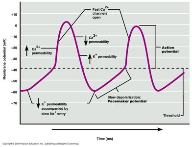
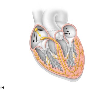
Pacemaker Cells: Autorhythmic cells generate action potentials, primarily at the sinoatrial (SA) node, which acts as the heart's pacemaker.
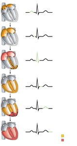
Sequence of Excitation: Electrical impulses travel from the SA node → AV node → AV bundle (His bundle) → right and left bundle branches → Purkinje fibers.
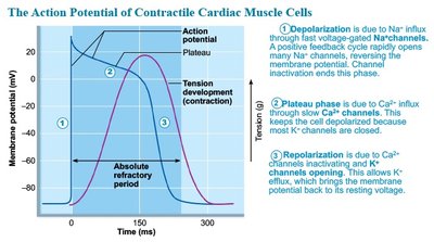
Action Potential Phases: Pacemaker potential (slow depolarization), depolarization (Ca2+ influx), repolarization (K+ efflux).
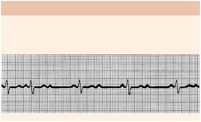
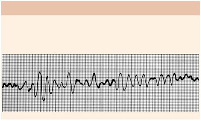
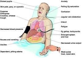
Clinical Relevance: Defects in the conduction system can cause arrhythmias, fibrillation, extrasystole, and heart block.
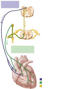
Extrinsic Innervation of the Heart
The heart's activity is modulated by the autonomic nervous system, which adjusts heart rate and force of contraction to meet the body's needs.
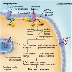
Cardioacceleratory Center: Sympathetic signals increase heart rate and contractility.
Cardioinhibitory Center: Parasympathetic signals (via the vagus nerve) decrease heart rate.
Clinical Relevance: Artificial pacemakers may be required if conduction defects prevent proper impulse transmission.
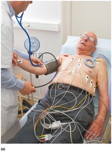
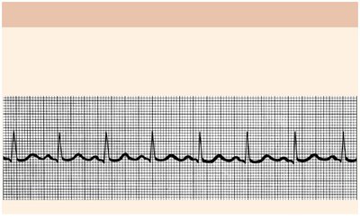
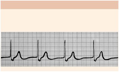
The Electrocardiogram (ECG)
An ECG is a graphical recording of the heart's electrical activity, used to diagnose conduction abnormalities and cardiac pathologies.
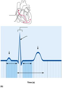
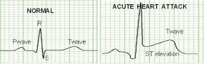
ECG Features: P wave (atrial depolarization), QRS complex (ventricular depolarization and atrial repolarization), T wave (ventricular repolarization).
Intervals: P-R interval, S-T segment, Q-T interval.
Clinical Relevance: Changes in ECG patterns can indicate ischemia, enlarged ventricles, or arrhythmias.
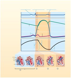
Mechanical Events of the Heart
The cardiac cycle consists of systole (contraction) and diastole (relaxation), with blood flow regulated by pressure changes and valve operation. Mechanical events follow electrical events observed on the ECG.
Phases of Cardiac Cycle: Ventricular filling, isovolumetric contraction, ventricular ejection, isovolumetric relaxation.
Valve Operation: AV valves close when ventricular pressure exceeds atrial pressure; SL valves open when ventricular pressure exceeds aortic pressure.
Heart Sounds: First sound (AV valves close), second sound (SL valves close).
Volumes: EDV (end diastolic volume), ESV (end systolic volume), SV (stroke volume).
Formula:
Regulation of Pumping (Heart Output)
Cardiac output (CO) is the amount of blood pumped by each ventricle per minute, determined by heart rate (HR) and stroke volume (SV). Regulation involves preload, contractility, and afterload.
Cardiac Output Formula:
Stroke Volume Formula:
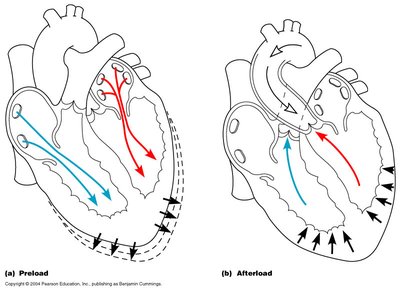
Preload: Degree of stretch of cardiac muscle before contraction; increased preload increases SV (Frank-Starling law).
Contractility: Contractile strength independent of muscle stretch; increased by sympathetic stimulation and positive inotropic agents.
Afterload: Pressure ventricles must overcome to eject blood; increased afterload decreases SV.
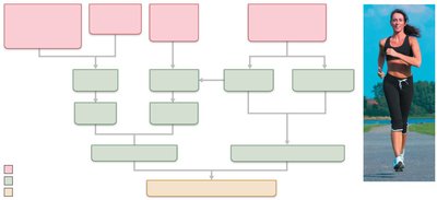
Regulation of Heart Rate: Influenced by autonomic nervous system, hormones, ions, age, gender, exercise, and temperature.

Developmental Aspects of the Heart
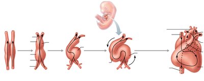
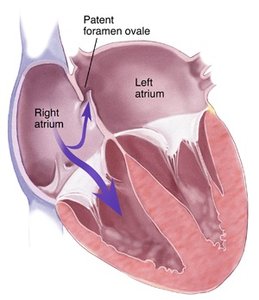
The heart develops from mesoderm, beginning as two endothelial tubes that fuse and undergo structural changes to form a four-chambered heart. Fetal heart structures, such as the foramen ovale and ductus arteriosus, bypass pulmonary circulation and close at birth.
Foramen Ovale: Connects atria; becomes fossa ovalis in adults.
Ductus Arteriosus: Connects pulmonary trunk to aorta; becomes ligamentum arteriosum in adults.
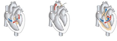
Congenital Heart Defects: Most common birth defects; include septal defects, coarctation of the aorta, and Tetralogy of Fallot.

Summary Table: Cardiac Cycle Phases and Associated Events
Phase | Electrical Event | Mechanical Event | Valve Status |
|---|---|---|---|
Ventricular Filling | P wave (atrial depolarization) | Atria contract, ventricles fill | AV open, SL closed |
Isovolumetric Contraction | QRS complex (ventricular depolarization) | Ventricles contract, all valves closed | AV closed, SL closed |
Ventricular Ejection | QRS complex | Blood ejected from ventricles | AV closed, SL open |
Isovolumetric Relaxation | T wave (ventricular repolarization) | Ventricles relax, all valves closed | AV closed, SL closed |
Key Terms and Definitions
Depolarization: The process by which cardiac cells become less negative, initiating contraction.
Repolarization: The return of cardiac cells to their resting negative state, leading to relaxation.
Arrhythmia: Abnormal heart rhythm.
Fibrillation: Rapid, irregular contractions; can be atrial or ventricular.
Stroke Volume (SV): Volume of blood ejected by a ventricle per beat.
Cardiac Output (CO): Volume of blood pumped by each ventricle per minute.
Preload: Degree of stretch of cardiac muscle before contraction.
Contractility: Strength of contraction at a given muscle length.
Afterload: Pressure the ventricles must overcome to eject blood.
