 Back
BackHemostasis: Mechanisms of Blood Clotting and Repair
Study Guide - Smart Notes
 Tailored notes based on your materials, expanded with key definitions, examples, and context.
Hemostasis
Overview of Hemostasis
Hemostasis is the physiological process that stops bleeding at the site of an injury. It involves a series of tightly regulated steps to prevent excessive blood loss while ensuring that blood flow is restored after repair. The process consists of three main phases: the vascular phase, the platelet phase, and the coagulation phase, followed by clot retraction and fibrinolysis.
Hemostasis: The cessation of bleeding.
Phases: Vascular phase, Platelet phase, Coagulation phase.
Additional steps: Clot retraction and Fibrinolysis.
Vascular Phase
Mechanisms of the Vascular Phase
The vascular phase is the initial response to blood vessel injury, characterized by a vascular spasm that reduces blood flow. This phase lasts about 30 minutes and involves several steps to minimize blood loss and prepare the site for further repair.
Endothelial cell contraction: Exposes the basement membrane to the bloodstream.
Release of chemical factors: ADP, tissue factor, prostacyclin, and endothelins are released to stimulate smooth muscle contraction and cell division.
Endothelial plasma membranes become sticky: This helps seal off blood flow.
Platelet Phase
Platelet Activation and Plug Formation
The platelet phase begins within 15 seconds after injury. Platelets adhere to exposed surfaces and aggregate to form a platelet plug, which temporarily seals small breaks in the vessel wall.
Platelet adhesion: Platelets attach to sticky endothelial surfaces, basement membranes, and exposed collagen fibers.
Platelet aggregation: Platelets stick together, forming a platelet plug.
Platelet Activation and Release of Compounds
Activated platelets release several compounds that promote clotting and tissue repair.
Adenosine diphosphate (ADP): Stimulates further platelet activation.
Thromboxane A2 and serotonin: Enhance vascular spasm and platelet aggregation.
Clotting factors: Essential for the coagulation phase.
Platelet-derived growth factor (PDGF): Promotes tissue repair.
Calcium ions: Required for several steps in coagulation.
Factors Limiting Platelet Plug Growth
Several mechanisms prevent the excessive growth of the platelet plug, ensuring that clotting is localized to the site of injury.
Prostacyclin: Inhibits platelet aggregation.
Inhibitory compounds from WBCs: Suppress platelet activity.
Circulating enzymes: Break down ADP.
Negative feedback from serotonin: Limits platelet activation.
Blood clot formation: Isolates the area of injury.
Coagulation Phase
Blood Clotting Mechanisms
The coagulation phase begins 30 seconds or more after injury. It involves a cascade of reactions that convert soluble fibrinogen into insoluble fibrin, forming a stable blood clot.
Cascade reactions: Chain reactions of enzymes and proenzymes.
Three pathways: Extrinsic, Intrinsic, and Common pathways.
Fibrin formation: Fibrinogen is converted to fibrin, which forms the clot.
Coagulation Pathways and Clotting Factors
Coagulation involves three pathways, each requiring specific clotting factors (procoagulants), which are proteins or ions in plasma.
Extrinsic pathway: Triggered by tissue damage outside the bloodstream.
Intrinsic pathway: Initiated by factors within the bloodstream.
Common pathway: Where both pathways converge to form the final clot.
Clotting factors: Essential for normal clotting.
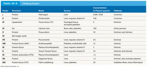
Clotting Factors Table
The following table summarizes the main clotting factors, their structure, source, concentration in plasma, and pathway involvement.
Factor | Structure | Name | Source | Concentration in Plasma (µg/mL) | Pathway |
|---|---|---|---|---|---|
I | Protein | Fibrinogen | Liver | 2500-3500 | Common |
II | Protein | Prothrombin | Liver, requires vitamin K | 100 | Common |
III | Protein | Tissue factor (TF) | Damaged tissues | Variable | Extrinsic and common |
IV | Ion | Calcium ions | Bone, diet, platelets | 5 | Extrinsic and intrinsic |
V | Protein | Proaccelerin | Liver, platelets | 10 | Extrinsic and intrinsic |
VII | Protein | Proconvertin | Liver, requires vitamin K | 0.5 | Extrinsic |
VIII | Protein | Antihemophilic factor | Platelets, endothelial cells | 1 | Intrinsic |
IX | Protein | Plasma thromboplastin factor | Liver, requires vitamin K | 1 | Intrinsic |
X | Protein | Stuart factor | Liver, requires vitamin K | 2 | Extrinsic and intrinsic |
XI | Protein | Plasma thromboplastin antecedent | Liver | 5 | Intrinsic |
XII | Protein | Hageman factor | Liver | 40 | Intrinsic, also activates fibrinolysis |
XIII | Protein | Fibrin stabilizing | Liver, platelets | 20 | Stabilizes fibrin, cross-links |
Extrinsic Pathway
The extrinsic pathway is activated when tissue outside the bloodstream is damaged. Damaged cells release tissue factor (TF), which interacts with other compounds to form an enzyme complex that activates Factor X.
Tissue factor (TF): Released by damaged cells.
Activation of Factor X: Initiates the common pathway.
Intrinsic Pathway
The intrinsic pathway begins with circulating proenzymes within the bloodstream. Activation occurs when collagen is exposed due to tissue damage, and platelets release factors such as PF-3. A series of reactions ultimately activates Factor X.
Collagen exposure: Triggers enzyme activation.
Platelet factors (PF-3): Released by platelets.
Activation of Factor X: Leads to the common pathway.
Common Pathway
The common pathway is where the intrinsic and extrinsic pathways converge. It results in the formation of the enzyme prothrombinase, which converts prothrombin to thrombin. Thrombin then converts fibrinogen to fibrin, forming the clot.
Prothrombinase: Enzyme formed at the convergence of pathways.
Conversion of prothrombin to thrombin:
Conversion of fibrinogen to fibrin:
Clot Retraction
Stabilization and Repair of the Injury Site
Clot retraction occurs once the fibrin meshwork has formed. Platelets and red blood cells adhere to the fibrin strands, and platelets contract to pull the torn edges of the vessel closer together. This reduces residual bleeding and stabilizes the injury site, facilitating tissue repair.
Platelet contraction: Pulls vessel edges together.
Reduces size of damaged area: Makes repair easier for fibrocytes, smooth muscle cells, and endothelial cells.
Duration: Continues over 30–60 minutes.
Fibrinolysis
Dissolution of the Clot
Fibrinolysis is the slow process of dissolving the clot after tissue repair is complete. Thrombin and tissue plasminogen activator (t-PA) activate plasminogen, which produces plasmin. Plasmin digests fibrin strands, removing the clot.
Activation of plasminogen: By thrombin and t-PA.
Plasmin: Enzyme that digests fibrin.
Removal of clot: Restores normal blood flow.
