 Back
BackHigher Mental Functions: Limbic System, Reticular Formation, EEG, Sleep, and Memory
Study Guide - Smart Notes
 Tailored notes based on your materials, expanded with key definitions, examples, and context.
Integrative Functions of the Limbic System and Reticular Formation
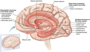
Limbic System: Structure and Function
The limbic system is a complex set of structures located on the medial aspect of each cerebral hemisphere and diencephalon. It is central to emotional processing, memory formation, and the regulation of autonomic and endocrine responses to emotional stimuli.
Amygdala: Involved in emotional reactions, especially fear processing.
Hippocampus: Essential for memory formation; damage can cause anterograde amnesia.
Parahippocampal Gyrus: Assists in memory processing as part of the hippocampal formation.
Cingulate Gyrus: Facilitates memory transfer and emotional regulation.
Hypothalamus: Maintains homeostasis and regulates the autonomic nervous system.
Anterior Thalamic Nuclei: Relay limbic influences to the cerebral cortex.
Fiber Tracts (Fornix, Anterior Commissure): Connect limbic structures for integrated function.
Example: The hippocampus is critical for forming new memories, as seen in patients with hippocampal damage who cannot form new long-term memories.
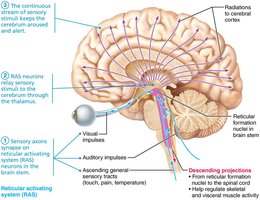
Reticular Formation: Significance and Connections
The reticular formation is a network of nuclei in the brainstem that influences arousal and consciousness. It projects to the thalamus and cerebral cortex, playing a crucial role in regulating wakefulness and alertness.
Ascending Pathways: Influence cortical arousal and consciousness.
Descending Pathways: Regulate skeletal and visceral muscle activity via the spinal cord.
Clinical Note: Inhibition by substances like opioids can suppress respiration and consciousness.
Sensory Pathways and Arousal via the Reticular Formation
Reticular Activating System (RAS) and Sensory Input
The reticular activating system (RAS) receives extensive sensory input, which is essential for maintaining brain arousal states and consciousness. Sensory axons synapse on RAS neurons, which relay information to the thalamus and then to the cerebral cortex.
Continuous Sensory Stream: Maintains cerebral alertness (e.g., smelling salts stimulate the RAS).
Thalamic Relay: Sensory data is projected to the cortex, modulating arousal levels.
Damage: Disruption can lead to coma or altered consciousness.
Example: The RAS is activated by sensory stimuli, which is why sudden noises can wake a sleeping person.
Understanding Electroencephalogram (EEG): Neuronal Synchrony and Brain Wave Patterns
Principles of EEG Recording
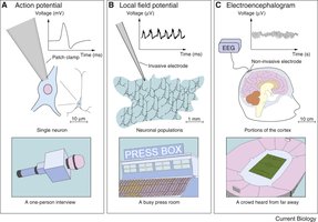
An electroencephalogram (EEG) records electrical activity from groups of neurons in the cerebral cortex using scalp electrodes. It measures postsynaptic potentials, not individual action potentials.
Signal Amplitude: EEG detects small electrical signals (microvolts) resulting from synchronized neuronal activity.
Types of Brain Waves:
Alpha Waves: Low amplitude, high frequency; relaxed, awake state.
Beta Waves: Lower amplitude, higher frequency; active thinking.
Theta Waves: Irregular; common in children, abnormal in adults.
Delta Waves: High amplitude, low frequency; deep sleep.
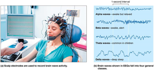
Example: Alpha waves are prominent when a person is awake but relaxed, while beta waves dominate during intense mental activity.
Sleep and Wakefulness: Stages, Regulation, and Functions
Sleep Stages and EEG Patterns
Sleep is divided into NREM (non-rapid eye movement) and REM (rapid eye movement) stages, each with distinct EEG patterns and physiological characteristics.
NREM Sleep: Four stages, progressing from light to deep sleep, characterized by increasing synchrony and amplitude of EEG waves.
REM Sleep: Follows NREM; EEG resembles wakefulness, dreaming occurs, and most skeletal muscles are inhibited.
Sleep Cycle: Alternates between NREM and REM throughout the night, each cycle lasting about 90 minutes.

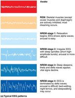
Example: REM sleep is associated with vivid dreams and muscle atonia, except for the eyes and diaphragm.
Regulation of Sleep and Circadian Rhythms
Sleep and wakefulness are regulated by the interplay of the RAS, thalamus, hypothalamic nuclei, and neurotransmitters such as orexin, histamine, and serotonin. The circadian rhythm aligns sleep-wake cycles with the day-night cycle.
Hypothalamic Role: Orexin-producing neurons are crucial for maintaining wakefulness; deficits can cause narcolepsy.
Active Sleep Regulation: Sleep is not just a passive process but involves active neural mechanisms.
The Impact of Sleep on Physiological Systems and Disorders
Consequences of Chronic Sleep Disruption
Chronic sleep loss can have widespread effects on health, including increased inflammation, reduced immune function, altered metabolism, and blood-brain barrier dysfunction.
Pro-inflammatory Response: Elevated cytokines (e.g., TNF-α, IL-6).
Metabolic Effects: Hyperglycemia, insulin resistance.
Cardiovascular and Neurological Impact: Increased stress, risk of neurological disorders.
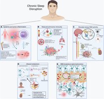
Example: Sleep apnea can lead to cardiovascular disease due to chronic intermittent hypoxia and sympathetic activation.
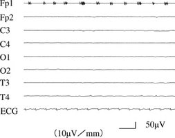
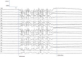
Clinical Relevance of EEG Findings
EEG in Diagnosis and Monitoring
EEG is a critical tool in clinical neurology for diagnosing epilepsy, assessing brain death, and monitoring brain activity in various conditions.
Brain Death: Flat EEG indicates absence of cortical activity, used in clinical death determination.
Epilepsy: Characteristic spike-and-wave patterns help classify seizure types and guide treatment.
Other Uses: Localizing brain lesions, monitoring anesthesia, and evaluating sleep disorders.
Hemispheric Lateralization in Language and Cognitive Processing
Functional Asymmetry of the Brain
The brain exhibits hemispheric lateralization, with the left hemisphere typically specializing in language, logic, and analytical tasks, while the right hemisphere excels in spatial, artistic, and emotional processing.
Language Centers: Broca's area (speech production) and Wernicke's area (comprehension) are usually in the left hemisphere.
Right Hemisphere: Handles nonverbal communication, emotional tone, and spatial awareness.
Corpus Callosum: Connects hemispheres, enabling integrated cognitive function.

Example: Damage to Broca's area results in expressive aphasia, while Wernicke's area damage causes receptive aphasia.
Memory: Pathways, Mechanisms, and Types
Stages and Structures of Memory
Memory involves the encoding, storage, and retrieval of information, with the hippocampus playing a central role in long-term memory formation.
Sensory Memory: Brief storage of sensory input.
Short-Term Memory (STM): Temporary holding of information; limited capacity and duration.
Long-Term Memory (LTM): Stable, enduring storage; involves structural changes in neural circuits.
Memory Consolidation: Influenced by excitement, rehearsal, and association.
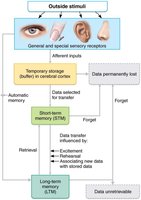
Example: Rehearsing a phone number helps transfer it from STM to LTM.
Types of Memory: Declarative vs. Procedural
Memory is divided into declarative (explicit) and procedural (implicit) types, each with distinct neural substrates.
Declarative Memory: Facts and events; conscious recall; hippocampus-dependent.
Procedural Memory: Skills and habits; unconscious recall; involves basal ganglia and cerebellum.
Example: Remembering historical dates (declarative) vs. riding a bicycle (procedural).
Cellular and Molecular Mechanisms of Long-Term Memory
Long-term memory formation involves synaptic plasticity, particularly long-term potentiation (LTP), which enhances synaptic strength through increased neurotransmitter release and receptor sensitivity.
NMDA Receptor: Allows Ca2+ influx after Mg2+ block is removed by strong stimulation, initiating LTP.
AMPA Receptor: Increased number/sensitivity augments synaptic response.
Protein Synthesis: Required for structural changes in synapses.
Neurogenesis: Occurs in the hippocampus, contributing to memory formation.
Equation (LTP Induction):
Example: Practicing a skill repeatedly strengthens the neural circuits involved, making the skill automatic over time.
