 Back
Histology and the Integumentary System: Structure and Function of Human Tissues
Study Guide - Smart Notes
 Tailored notes based on your materials, expanded with key definitions, examples, and context.
Overview of Tissues and Histology
Definition and Classification of Tissues
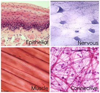
Tissues are groups of cells that are similar in structure and perform common functions. The study of tissues and their arrangement into organs is known as histology. The human body is composed of four primary tissue types: epithelial, connective, muscle, and nervous tissue.
Epithelial Tissue: Covers surfaces, lines cavities, and forms glands.
Connective Tissue: Supports, binds, and protects other tissues.
Muscle Tissue: Responsible for movement.
Nervous Tissue: Conducts electrical impulses and processes information.
Epithelial Tissue
Classification by Cell Shape
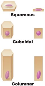
Epithelial tissues are classified based on cell shape:
Squamous: Scale-like, flat cells.
Cuboidal: Cube-shaped cells.
Columnar: Tall, column-like cells.
Classification by Cell Organization
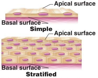
Epithelial tissues are also classified by the number of cell layers:
Simple: One layer of cells.
Stratified: Two or more layers of cells.
Pseudostratified: Appears layered but is actually a single layer.
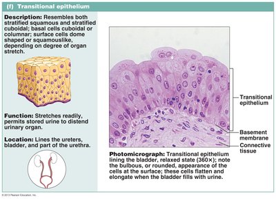
Transitional: Can change shape; dome-shaped when relaxed, squamous when stretched.
Major Functions of Epithelial Tissue
Protection: Shields underlying tissues from injury and pathogens.
Absorption: Takes in nutrients and substances.
Filtration: Filters substances passing through tissues.
Excretion: Removes waste products.
Secretion: Produces and releases substances such as mucus and hormones.
Sensory Reception: Detects environmental changes.
Types of Epithelial Tissue
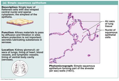
Simple Squamous Epithelium
Single layer of flat cells; allows materials to pass by diffusion and filtration. Found in air sacs of lungs, lining of blood vessels, and serous membranes.
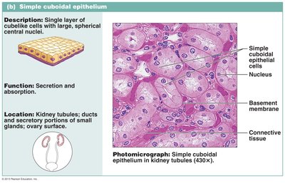
Simple Cuboidal Epithelium
Single layer of cube-shaped cells; functions in secretion and absorption. Located in kidney tubules and ducts of glands.
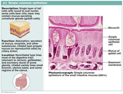
Simple Columnar Epithelium
Single layer of tall cells; specialized for absorption and secretion. Found in digestive tract lining and some glands.
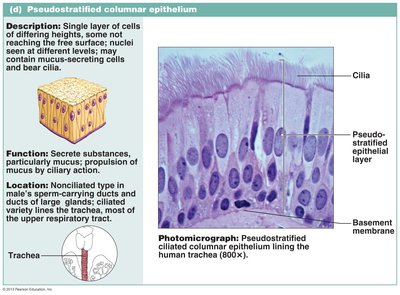
Pseudostratified (Ciliated) Columnar Epithelium
Single layer of cells of varying heights; often ciliated and involved in secretion and movement of mucus. Located in the trachea and upper respiratory tract.
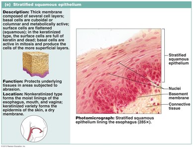
Stratified Squamous Epithelium
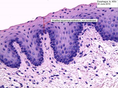
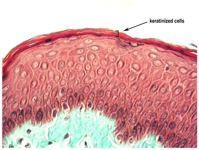
Multiple layers of cells; protects against abrasion. Non-keratinized forms line moist surfaces (e.g., esophagus), while keratinized forms are found in the skin.
Stratified Cuboidal Epithelium
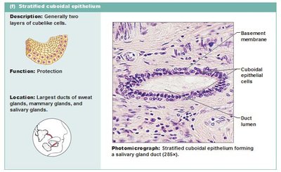
Generally two layers of cube-shaped cells; provides protection. Found in ducts of sweat glands and salivary glands.
Stratified Columnar Epithelium
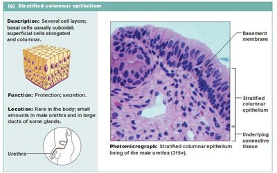
Several cell layers; basal cells are usually cuboidal, superficial cells are columnar. Functions in protection and secretion. Rare, found in male urethra and large ducts.
Transitional Epithelium
Resembles both stratified squamous and cuboidal; stretches readily and permits distension of urinary organs. Found in the bladder, ureters, and part of the urethra.
Keratin and Epithelial Specializations
Keratinized vs. Non-Keratinized Epithelium
Keratin is a family of fibrous structural proteins that assemble into bundles to form intermediate filaments. Keratinized epithelium forms a tough, dehydrated outer layer, protecting against abrasion and dehydration. Non-keratinized epithelium must be kept moist by bodily secretions.
Connective Tissue
Classification and Functions
Connective tissue (CT) is the most abundant and widely distributed tissue in the body. It is classified into four main classes:
Connective tissue proper: Loose (areolar, adipose, reticular) and dense (regular, irregular, elastic).
Cartilage: Hyaline, elastic, fibrocartilage.
Bone: Compact and spongy.
Blood: Contains RBCs, WBCs, and platelets.
Major functions include binding and support, protection, insulation, storing reserve fuel, and transporting substances.
Muscle Tissue
Types and Functions
Muscle tissue is specialized for contraction and movement. There are three types:
Skeletal Muscle: Supports and moves the body; voluntary control.
Cardiac Muscle: Contracts rhythmically to propel blood; found in the heart.
Smooth Muscle: Responsible for contractility of hollow organs; involuntary control.
Nervous Tissue
Structure and Function
Nervous tissue consists of two major cell types:
Neurons: Specialized cells that generate and conduct nerve impulses.
Neuroglia: Supporting cells that insulate, protect, and support neurons.
Integumentary System
Structure of Skin
The skin is composed of two distinct regions:
Epidermis: Superficial layer made of keratinized stratified squamous epithelium. Thin skin has four layers; thick skin has five layers.
Dermis: Deep layer of fibrous connective tissue attached to the epidermis by a basement membrane. Supports the epidermis and connects it to underlying fat tissue (hypodermis).
Dermal Layers
Reticular Layer: Dense irregular connective tissue; contains glands, nerves, and blood vessels. Responsible for primary sensory functions.
Papillary Layer: Loose areolar connective tissue; adheres to the epidermis.
Summary Table: Epithelial Tissue Types
Type | Cell Shape | Layers | Function | Location |
|---|---|---|---|---|
Simple Squamous | Flat | 1 | Diffusion, filtration | Air sacs of lungs, blood vessels |
Simple Cuboidal | Cube | 1 | Secretion, absorption | Kidney tubules, glands |
Simple Columnar | Tall | 1 | Absorption, secretion | Digestive tract |
Pseudostratified Columnar | Tall | 1 (appears layered) | Secretion, movement of mucus | Trachea, upper respiratory tract |
Stratified Squamous | Flat | Multiple | Protection | Skin, esophagus, mouth |
Stratified Cuboidal | Cube | 2 | Protection | Sweat glands, salivary glands |
Stratified Columnar | Tall | Multiple | Protection, secretion | Male urethra, large ducts |
Transitional | Dome-shaped/squamous | Multiple | Stretching | Bladder, ureters |
Example: The lining of the urinary bladder is transitional epithelium, allowing it to stretch as the bladder fills.
Example: The skin's outer layer is keratinized stratified squamous epithelium, providing protection against dehydration and abrasion.
Additional info: The notes above expand on brief points with academic context, definitions, and examples to ensure completeness and clarity for exam preparation.
