 Back
BackHistology: Epithelial Tissue Structure and Classification
Study Guide - Smart Notes
 Tailored notes based on your materials, expanded with key definitions, examples, and context.
Tissue Organization
Introduction to Tissues
Tissues are groups of similar cells and extracellular material that perform a common function in the body. The study of tissues is called histology. There are four primary tissue types: epithelial, connective, muscle, and nervous tissue. Each type varies in structure, function, and the composition of the extracellular matrix.
Epithelial Tissue
General Characteristics
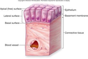
Epithelial tissue (or epithelium) consists of one or more layers of closely packed cells with minimal extracellular matrix. It covers body surfaces, lines body cavities and organ cavities, and forms the majority of glands.
Cellularity: Composed almost entirely of tightly packed cells.
Polarity: Has an apical (free) surface, lateral surfaces with intercellular junctions, and a basal surface attached to connective tissue.
Attachment to Basement Membrane: The basal surface is attached to a basement membrane, which is a selective barrier composed of collagen fibers, glycoproteins, and carbohydrates.
Avascularity: Lacks blood vessels; nutrients diffuse from underlying tissues.
Extensive Innervation: Contains many nerve endings for sensory detection.
High Regeneration Capacity: Frequently damaged or lost cells are replaced by mitosis of stem cells near the basement membrane.
Functions of Epithelial Tissue
Physical Protection: Shields underlying tissues from dehydration, abrasion, and destruction by physical, chemical, or biological agents.
Selective Permeability: Acts as a gatekeeper, allowing certain substances to pass while blocking others.
Secretions: Forms exocrine and endocrine glands that produce and release substances such as sweat, hormones, and enzymes.
Sensations: Contains nerve endings that detect stimuli such as touch, pressure, temperature, and pain.
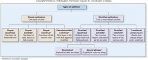
Classification of Epithelial Tissue
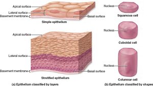
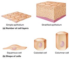
By Number of Cell Layers and Cell Shape
Epithelial tissues are classified based on the number of cell layers and the shape of the cells at the apical surface.
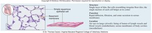
Simple epithelium: One layer of cells; all cells touch the basement membrane.
Stratified epithelium: Two or more layers; only the deepest layer touches the basement membrane.
Pseudostratified epithelium: Appears layered but all cells touch the basement membrane.
Squamous cells: Flat and thin.
Cuboidal cells: Cube-shaped, as tall as they are wide.
Columnar cells: Taller than they are wide.
Transitional cells: Change shape from round to flat when stretched (found in the urinary tract).
Examples of Epithelial Tissue Types
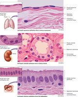
Simple squamous epithelium: Single layer of flat cells; allows rapid diffusion and filtration. Found in alveoli of lungs, lining of blood vessels, and serous membranes.
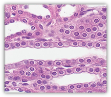
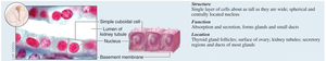
Simple cuboidal epithelium: Single layer of cube-shaped cells; functions in absorption and secretion. Found in kidney tubules and glands.
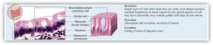
Simple columnar epithelium: Single layer of tall, column-shaped cells; functions in absorption and secretion, often contains goblet cells. Found in the digestive tract lining.
Stratified squamous epithelium: Multiple layers; protects against abrasion. Keratinized type forms the epidermis of skin; nonkeratinized type lines the mouth, esophagus, and vagina.
Stratified cuboidal and columnar epithelium: Rare; found in some glands and ducts.
Pseudostratified columnar epithelium: Appears layered but all cells touch the basement membrane; often ciliated. Found in the respiratory tract.
Transitional epithelium: Multiple layers of cells that change shape; found in the urinary bladder.

Summary Table: Types of Epithelia
Type | Structure | Function | Location |
|---|---|---|---|
Simple squamous | Single layer, flat cells | Diffusion, filtration | Alveoli, blood vessels, serous membranes |
Simple cuboidal | Single layer, cube-shaped cells | Absorption, secretion | Kidney tubules, glands |
Simple columnar | Single layer, tall cells | Absorption, secretion | Digestive tract lining |
Stratified squamous | Multiple layers, flat cells at surface | Protection | Skin, mouth, esophagus, vagina |
Pseudostratified columnar | Single layer, appears multilayered | Secretion, movement of mucus | Respiratory tract |
Transitional | Multiple layers, shape varies | Stretching, distension | Urinary bladder |
