 Back
BackHistology of Human Tissues: Epithelial, Connective, Muscle, and Nervous Tissue
Study Guide - Smart Notes
 Tailored notes based on your materials, expanded with key definitions, examples, and context.
Histology: The Study of Tissues
Histology is the microscopic study of tissues, which are groups of cells that perform specific functions. Understanding tissue types is fundamental to anatomy and physiology, as tissues form the structural and functional basis of organs and systems.
Epithelial Tissue (ET)
Overview of Epithelial Tissue
Epithelial tissue covers body surfaces, lines internal cavities, and forms glands. It is specialized for protection, absorption, secretion, and filtration.
Key Characteristics: Closely packed cells, minimal extracellular matrix, avascular, high regenerative capacity.
Functions: Protection, absorption, secretion, filtration.
Types of Epithelial Tissue
Simple Squamous Epithelium: Location: Lung alveoli, serous membranes, capillaries Function: Diffusion, secretion, reduces friction Example: Gas exchange in alveoli
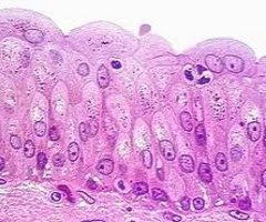
Stratified Squamous Epithelium: Location: Epidermis, lining of mouth, esophagus, vagina, anal canal Function: Protection from abrasion, pathogens, chemical attack Example: Skin protects against mechanical injury
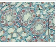
Simple Cuboidal Epithelium: Location: Kidney tubules, glands Function: Secretion, absorption Example: Kidney tubules filter blood
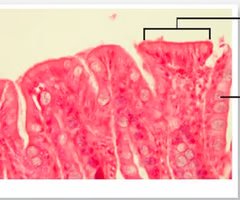
Simple Columnar Epithelium: Location: Lining of digestive tract (stomach to rectum) Function: Secretion, absorption Example: Absorption of nutrients in intestines
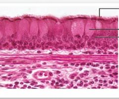
Pseudostratified Columnar Epithelium (with cilia and goblet cells): Location: Nasal cavity, trachea, bronchi Function: Secrete and propel mucus Example: Cilia move mucus in respiratory tract
Transitional Epithelium: Location: Ureters, urinary bladder Function: Distension/stretching Example: Bladder expands to store urine
Connective Tissue (CT)
Overview of Connective Tissue
Connective tissue supports, binds, and protects other tissues and organs. It is characterized by abundant extracellular matrix and diverse cell types.
Key Components: Cells (fibroblasts, adipocytes, chondrocytes, osteocytes), fibers (collagen, elastic, reticular), ground substance.
Functions: Support, protection, transport, insulation, energy storage.
Fibrous Connective Tissue (Proper)
Loose Areolar Connective Tissue: Location: Hypodermis, abdomen, breasts Function: Insulation, cushion, energy storage Example: Cushions organs beneath skin
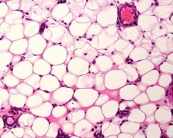
Loose Adipose Connective Tissue: Location: Hypodermis, abdomen, breasts Function: Insulation, cushion, energy storage Example: Fat storage in adipocytes
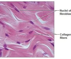
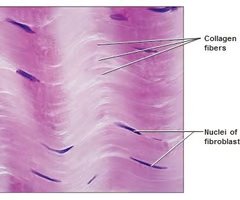
Dense Regular Connective Tissue: Location: Tendons, ligaments Function: Strong attachment in one direction Example: Tendons connect muscle to bone
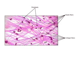
Dense Irregular Connective Tissue: Location: Dermis of skin Function: Strong attachment in different directions Example: Dermis resists multidirectional stress
Other Connective Tissues
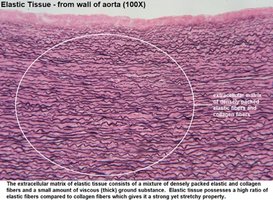
Dense Elastic Connective Tissue: Location: Vocal cords, wall of aorta Function: Recoil Example: Aorta stretches and recoils with blood flow
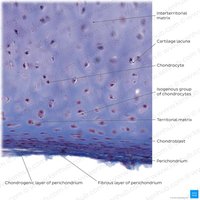
Hyaline Cartilage: Location: Ends of long bones, embryonic skeleton, nose, trachea Function: Flexible support, reduces friction Example: Articular cartilage in joints
Fibrocartilage: Location: Intervertebral discs, pubic symphysis, meniscus Function: Shock absorber; prevents bone-to-bone contact Example: Intervertebral discs absorb spinal shock
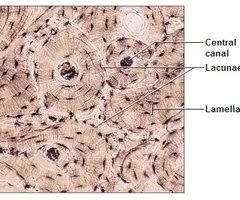
Osseous Tissue (Bone): Location: Axial & appendicular skeleton Function: Protection, support, movement, hematopoiesis Example: Bone marrow produces blood cells
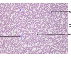
Blood: Location: Cardiovascular system Function: Transport nutrients & wastes; immunity Example: Red blood cells carry oxygen
Muscle Tissue (MT)
Overview of Muscle Tissue
Muscle tissue is specialized for contraction, enabling movement and force generation. It is classified by structure and function.
Key Types: Smooth, cardiac, skeletal
Functions: Movement, posture, heat production, blood flow
Types of Muscle Tissue
Smooth Muscle: Location: Walls of hollow organs (stomach, intestines), arteries, veins Function: Involuntary contractions (peristalsis) Example: Moves food through digestive tract
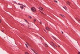
Cardiac Muscle: Location: Heart Function: Involuntary contractions to pump blood Example: Heartbeat
Skeletal Muscle: Location: Attached to skeleton & facial tissue Function: Voluntary contraction; thermoregulation; shivering Example: Movement of limbs
Nervous Tissue
Overview of Nervous Tissue
Nervous tissue is specialized for communication via electrical and chemical signals. It forms the brain, spinal cord, and peripheral nerves.
Key Cells: Neurons (transmit signals), neuroglia (support neurons)
Functions: Sensory input, integration, motor output
Summary Table: Tissue Types and Functions
Tissue Type | Location | Function |
|---|---|---|
Simple Squamous Epithelium | Lung alveoli, capillaries | Diffusion, secretion |
Stratified Squamous Epithelium | Epidermis, mouth | Protection |
Simple Cuboidal Epithelium | Kidney tubules, glands | Secretion, absorption |
Simple Columnar Epithelium | Digestive tract | Absorption, secretion |
Pseudostratified Columnar Epithelium | Trachea, bronchi | Mucus secretion |
Transitional Epithelium | Bladder, ureters | Stretching |
Loose Areolar CT | Hypodermis | Cushion, insulation |
Adipose CT | Abdomen, breasts | Energy storage |
Dense Regular CT | Tendons, ligaments | Attachment |
Dense Irregular CT | Dermis | Strength |
Dense Elastic CT | Aorta | Recoil |
Hyaline Cartilage | Joints, nose | Support |
Fibrocartilage | Discs, meniscus | Shock absorption |
Osseous Tissue | Skeleton | Support, protection |
Blood | Vessels | Transport |
Smooth Muscle | Organs | Involuntary movement |
Cardiac Muscle | Heart | Pumping blood |
Skeletal Muscle | Skeleton | Voluntary movement |
Nervous Tissue | Brain, nerves | Communication |
Additional info: Nervous tissue details were inferred for completeness, as the original file focused on epithelial, connective, and muscle tissues.
