 Back
Histology: Structure and Function of Tissues
Study Guide - Smart Notes
 Tailored notes based on your materials, expanded with key definitions, examples, and context.
Histology: The Study of Tissues
Definition and Overview
Histology is the branch of biology that examines tissues, which are groups of related cells working together to perform specific functions. Each tissue consists of cellular components and an extracellular matrix (ECM), which provides structural and functional support.
Tissue: Group of related cells performing common functions.
Extracellular Matrix (ECM): Material surrounding cells, composed of ground substance and protein fibers.
Extracellular Fluid (ECF): Fluid component within the ECM.
Four Primary Tissue Types
Epithelial Tissue
Epithelial tissue forms tightly packed sheets of cells with minimal ECM, covering and lining body surfaces and forming glands.
Structure: Tightly packed sheets of cells.
Function: Protection, secretion, absorption, and sensation.
Location: Covers body surfaces, lines cavities, forms glands.
Connective Tissue
Connective tissues are characterized by scattered cells within a prominent ECM, connecting, supporting, protecting, and transporting substances throughout the body.
Structure: Cells scattered in ECM.
Function: Binding, support, protection, transport.
Location: Connects all other tissues.
Muscle Tissue
Muscle tissue consists of specialized cells capable of contraction, generating force and movement.
Structure: Contractile cells (myocytes).
Function: Movement and force generation.
Nervous Tissue
Nervous tissue is composed of neurons and supporting cells, specialized for rapid communication.
Structure: Neurons and neuroglial cells.
Function: Generation, transmission, and reception of signals.
Extracellular Matrix (ECM)
Composition and Function
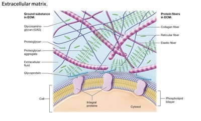
The ECM is a liquid, gel, or solid substance surrounding tissue cells, built from ground substance and protein fibers. It provides strength, directs cell placement, regulates cell activity, and holds cells together.
Ground Substance: Gel-like fluid with water, ions, nutrients, and macromolecules.
Protein Fibers: Collagen, elastic, and reticular fibers.
Ground Substance Macromolecules
Glycosaminoglycans (GAGs): Long polysaccharide chains creating osmotic gradients to resist compression.
Proteoglycans: GAGs bound to protein cores, forming aggregates for firmness and protection.
Glycoproteins: Cell-adhesion molecules anchoring cells to ECM and each other.
Protein Fibers in ECM
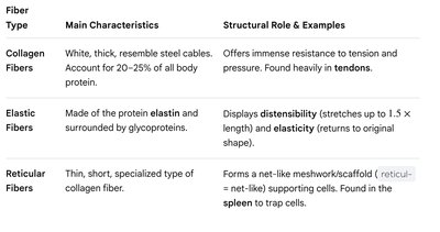
Protein fibers provide structural support and elasticity to tissues.
Collagen Fibers: White, thick, steel cable-like; resist tension and pressure; found in tendons.
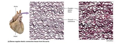
Elastic Fibers: Made of elastin; stretch and recoil; found in skin, lungs, blood vessels.
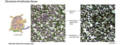
Reticular Fibers: Thin, net-like; support cells; found in spleen, lymph nodes.
Cell Junctions
Types and Functions
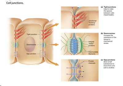
Cell junctions are specialized structures connecting adjacent cells, maintaining tissue integrity and communication.
Tight Junctions: Seal spaces between cells, making tissue impermeable.
Desmosomes: Distribute mechanical stress, allow fluid passage, attached to intermediate filaments.
Gap Junctions: Protein channels allowing rapid communication and substance flow between cells.
Structure and Components of Epithelial Tissue
Structural Characteristics
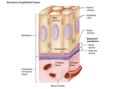
Epithelial tissue is organized in sheets of closely packed cells with minimal ECM, interconnected by cell junctions. It exhibits polarity with distinct apical and basal surfaces, anchored by a basement membrane.
Cell Arrangement: Sheets of closely packed cells.
Cell Junctions: Tight junctions and desmosomes.
Polarity: Apical (free) and basal (attached) surfaces.
Basement Membrane: Basal lamina (epithelial origin) and reticular lamina (connective origin).
Classification of Epithelia
Criteria for Classification
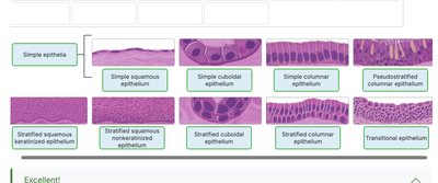
Epithelia are classified by the number of cell layers and the shape of cells.
Number of Layers: Simple (one layer), stratified (multiple layers), pseudostratified (appears layered, but all cells touch basement membrane).
Cell Shape: Squamous (flat), cuboidal (cube-like), columnar (tall).
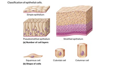
Types of Simple Epithelium
Simple Squamous and Cuboidal Epithelium
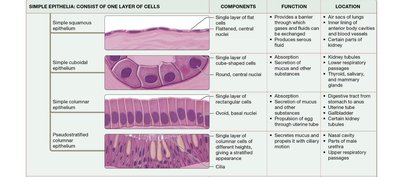
Simple epithelia consist of a single cell layer, facilitating rapid diffusion and secretion.
Simple Squamous: Flat cells; rapid diffusion; found in lungs, blood vessels.
Simple Cuboidal: Cube-shaped cells; rapid diffusion and secretion; found in kidney tubules, thyroid gland.
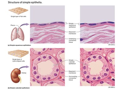
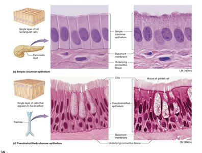
Simple Columnar and Pseudostratified Epithelium
Simple Columnar: Tall, rectangular cells; absorption and secretion; found in digestive tract.
Pseudostratified Columnar: Appears layered; all cells touch basement membrane; contains goblet cells; found in respiratory passages.
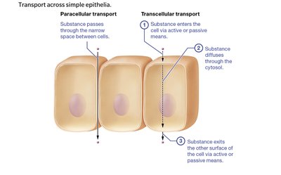
Transport Across Simple Epithelia
Paracellular vs. Transcellular Transport
Substances cross simple epithelia via two main pathways: paracellular (between cells) and transcellular (through cells).
Paracellular Transport: Limited by tight junctions; substances pass between cells.
Transcellular Transport: Substances enter, diffuse through cytosol, and exit the cell.
Types of Stratified Epithelium
Stratified Squamous, Cuboidal, Columnar, and Transitional Epithelium
Stratified epithelia consist of multiple cell layers, providing protection and specialized functions.
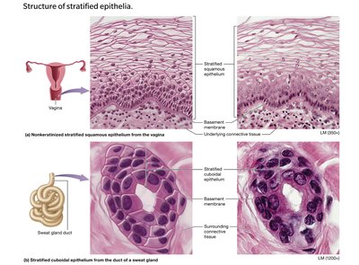
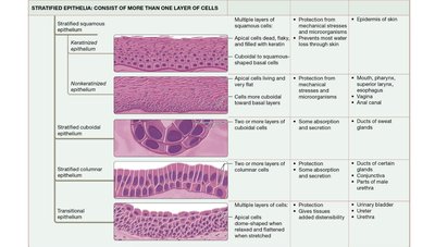
Stratified Squamous: Keratinized (skin) and nonkeratinized (moist linings).
Stratified Cuboidal: Two layers; lines sweat gland ducts.
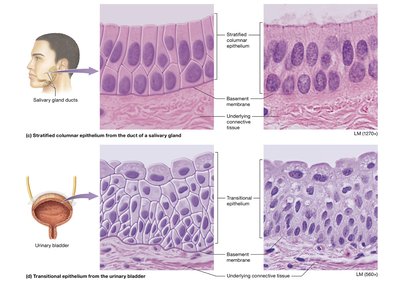
Stratified Columnar: Tall cells atop cuboidal cells; lines ducts, male urethra, conjunctiva.
Transitional: Dome-shaped apical cells; stretches; found in urinary system.
Summary Tables: Epithelial Types
Simple Epithelia
Type | Components | Function | Location |
|---|---|---|---|
Simple squamous | Single layer, flat cells | Rapid diffusion | Lungs, blood vessels |
Simple cuboidal | Single layer, cube cells | Absorption, secretion | Kidney tubules, thyroid |
Simple columnar | Single layer, tall cells | Absorption, secretion | Digestive tract |
Pseudostratified columnar | Single layer, nuclei at different heights | Mucus secretion, protection | Respiratory passages |
Stratified Epithelia
Type | Components | Function | Location |
|---|---|---|---|
Keratinized stratified squamous | Multiple layers, dead apical cells | Protection | Skin |
Nonkeratinized stratified squamous | Multiple layers, living apical cells | Protection | Mouth, esophagus, vagina |
Stratified cuboidal | Two layers, cube cells | Protection, secretion | Sweat glands |
Stratified columnar | Multiple layers, tall cells | Protection, secretion | Salivary glands, urethra |
Transitional | Multiple layers, dome-shaped apical cells | Distensibility | Urinary bladder |
Carcinogens and Epithelial Tissues
Carcinomas
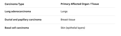
Carcinomas are cancers originating from epithelial tissues, which are highly susceptible due to rapid cell division and environmental exposure.
Carcinoma Type | Primary Affected Organ / Tissue |
|---|---|
Lung adenocarcinoma | Lungs |
Ductal and papillary carcinoma | Breast tissue |
Basal cell carcinoma | Skin (epithelial layers) |
Glands and Secretion
Exocrine and Endocrine Glands
Glands are classified by their method of secretion and structural complexity.
Exocrine Glands: Secrete products via ducts onto surfaces; local action.
Endocrine Glands: Secrete hormones directly into bloodstream; systemic action.
Unicellular and Multicellular Exocrine Glands
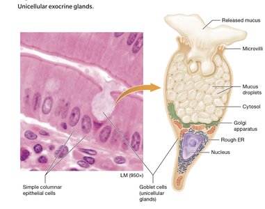
Unicellular: Goblet cells secrete mucus in digestive and respiratory tracts.
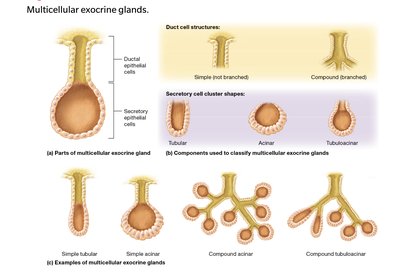
Multicellular: Complex glands with branching ducts and secretory units.
Modes of Exocrine Secretion
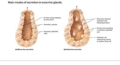
Merocrine: Products released by exocytosis; cell remains intact.
Holocrine: Products released when cell ruptures and dies; cell replaced by mitosis.
Apocrine: Apical portion pinched off; cell survives and repairs.
Connective Tissue Proper
Cells of Connective Tissue Proper
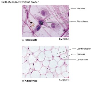
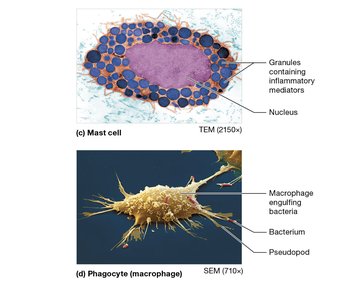
Connective tissue proper contains resident and migrant cells, each with specialized functions.
Fibroblasts: Synthesize protein fibers and ECM.
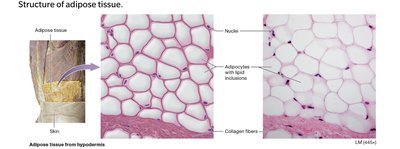
Adipocytes: Store fat; nucleus pushed to cell edge.
Mast Cells: Contain granules with inflammatory mediators.
Phagocytes: Ingest foreign invaders and debris.
Types of Connective Tissue Proper
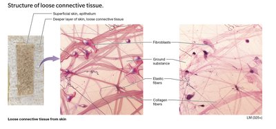
Loose Connective Tissue: Soft, jelly-like ECM; supports epithelium; rich in blood supply.
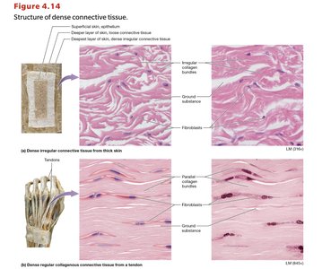
Dense Connective Tissue: High density of protein fibers; structural strength.
Reticular Tissue: Web-like network; traps cells; found in lymph nodes, spleen.
Adipose Tissue: Fat storage; insulation, protection, energy reserve.
Summary Table: Connective Tissue Proper
Type | Main Components | Function | Location |
|---|---|---|---|
Loose connective tissue | Ground substance, fibroblasts, adipocytes | Support, nutrient supply, immune defense | Under skin, lining organs |
Dense irregular | Collagen fibers, random arrangement | Resists tension in all directions | Dermis, organ capsules |
Dense regular collagenous | Collagen fibers, parallel arrangement | Strength in one direction | Tendons, ligaments |
Dense regular elastic | Elastic fibers, parallel arrangement | Stretch and recoil | Aorta, spinal ligaments |
Reticular tissue | Reticular fibers, reticular cells | Trap cells, support | Lymph nodes, spleen |
Adipose tissue | Adipocytes, minimal ECM | Energy storage, insulation, protection | Under skin, around organs |
 *Additional info: Expanded explanations, tables, and academic context were added to ensure completeness and clarity for college-level study.*
