 Back
BackHistology: Structure and Function of Tissues
Study Guide - Smart Notes
 Tailored notes based on your materials, expanded with key definitions, examples, and context.
Histology and Types of Tissues
Introduction to Tissues
Histology is the study of the normal structure of tissues, which are groups of structurally and functionally related cells and their external environment that together perform common functions. All tissues share two basic components: a discrete population of cells related in structure and function, and the surrounding material called the Extracellular Matrix (ECM).
Tissue: Group of cells and their environment performing a common function.
Histology: Study of tissue structure.
Extracellular Matrix (ECM): Material surrounding cells, varies by tissue type.
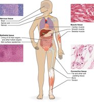
Types of Tissues
There are four primary tissue types in the human body, each with distinct functions and structural characteristics:
Epithelial Tissue: Sheets of tightly packed cells with little ECM; covers and lines body surfaces and cavities, forms parts of glands.
Connective Tissue: Connects other tissues; cells are scattered through ECM; binds, supports, protects, and allows transport of substances.
Muscular Tissue: Cells contract and generate force; little ECM.
Nervous Tissue: Cells (neurons) generate, send, and receive messages; includes supporting cells with unique ECM.
Extracellular Matrix (ECM)
Components and Functions
The ECM is composed of substances surrounding the cells in a tissue, providing strength, directing cell placement, regulating development, and holding cells in position. It consists of ground substance and protein fibers.
Ground Substance: Gel-like, contains extracellular fluid (ECF), water, ions, nutrients, and macromolecules.
Protein Fibers: Provide tensile strength; types include collagen, elastic, and reticular fibers.
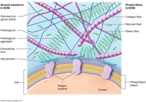
Ground Substance
Glycosaminoglycans (GAGs): Negatively charged polysaccharides; attract ions and water.
Proteoglycans: GAGs bonded to protein core; form aggregates, resist compression, act as diffusion barriers.
Glycoproteins (Cell-Adhesion Molecules, CAMs): Bind cell surface proteins and fibers, maintain tissue architecture.
Protein Fibers
Collagen Fibers: Most abundant, resistant to tension and pressure.
Elastic Fibers: Made of elastin, stretch and return to original length.
Reticular Fibers: Thinner collagen, form scaffolds and webs in organs.
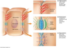
Cell Junctions
Types of Cell Junctions
Cell junctions are connections between neighboring cells, linked by integral proteins. They are essential for tissue integrity and communication.
Tight (Occluding) Junctions: Integral "locking" proteins prevent passage of macromolecules; some are leaky.
Desmosomes: "Linker" proteins distribute mechanical stress.
Gap Junctions: Protein channels allow small substances to pass freely between cells.
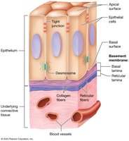
Epithelial Tissues
Functions of Epithelial Tissue
Epithelial tissue is found on every external and internal body surface, acting as a barrier between the body and the environment. Its functions include:
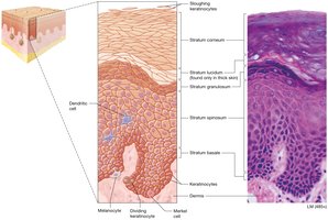
Protection: Shields underlying tissue, produces keratin, rapid mitosis.
Immune Defenses: Contains immune cells.
Secretion: Forms glands producing sweat, oil, hormones.
Transport: Selectively permeable barriers for substance movement.
Sensation: Supplied with nerves, detects environmental changes.
Components of Epithelia
The ECM of epithelia is located beneath the cells in the basement membrane, anchoring the tissue to underlying connective tissue. The basement membrane has two components:
Basal Lamina: ECM of epithelial tissue; collagen fibers and ground substance.
Reticular Lamina: Produced by connective tissue; reticular fibers and ground substance.
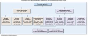
Classification of Epithelia
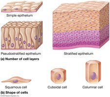
Epithelia are classified by the number of cell layers and the shape of the cells:
Simple Epithelia: Single layer of cells.
Stratified Epithelia: More than one layer of cells.
Pseudostratified Epithelia: Single layer that appears multilayered.
Squamous Cells: Flattened.
Cuboidal Cells: Short, square.
Columnar Cells: Tall, elongated.
Covering and Lining Epithelia
Simple Epithelia
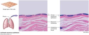
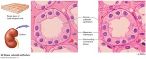
Simple epithelia are one cell layer thick and are found lining hollow organs and surfaces where diffusion or transport occurs.
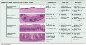
Simple Squamous: Flat cells, rapid diffusion; found in air sacs of lungs, serous membranes, blood vessels.
Simple Cuboidal: Cube-shaped cells, diffusion and secretion; found in kidney tubules and glands.
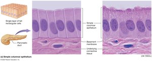
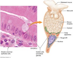
Simple Columnar: Tall cells, absorption and secretion; found in small intestine, uterine tube, kidney tubules.
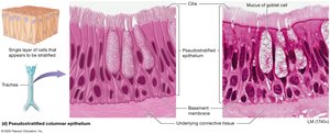
Pseudostratified Columnar: Appears stratified, ciliated, contains goblet cells; found in respiratory passages.
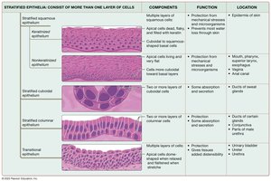
Stratified Epithelia
Stratified epithelia are thicker and provide protection in areas of high stress. Cell shape changes throughout the thickness, named by apical layer shape.
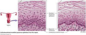
Stratified Squamous: Keratinized (skin) and nonkeratinized (mouth, esophagus, vagina).
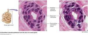
Stratified Cuboidal: Rare, lines ducts of sweat glands.
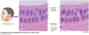
Stratified Columnar: Rare, found in salivary gland ducts, male urethra, conjunctiva.
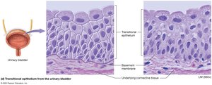
Transitional: Dome-shaped apical cells, stretchable; found in urinary bladder, ureters.
Summary Tables of Epithelial Tissues
The following tables summarize the components, functions, and locations of simple and stratified epithelia:
Type | Components | Function | Location |
|---|---|---|---|
Simple Squamous | Single layer, flat cells | Diffusion, filtration | Lungs, blood vessels |
Simple Cuboidal | Single layer, cube-shaped | Absorption, secretion | Kidney tubules, glands |
Simple Columnar | Single layer, tall cells | Absorption, secretion | Digestive tract, uterine tubes |
Pseudostratified Columnar | Single layer, ciliated | Secretion, movement | Respiratory tract |
Type | Components | Function | Location |
|---|---|---|---|
Keratinized Stratified Squamous | Multiple layers, dead apical cells | Protection | Skin |
Nonkeratinized Stratified Squamous | Multiple layers, living apical cells | Protection | Mouth, esophagus, vagina |
Stratified Cuboidal | Two layers, cube-shaped | Protection, secretion | Sweat gland ducts |
Stratified Columnar | Few layers, columnar apical | Protection, secretion | Salivary gland ducts |
Transitional | Multiple layers, dome-shaped apical | Stretching | Urinary bladder |
Glandular Epithelia
Types of Glands
Glands are structures that make and secrete products, arising from epithelial tissue. They release products by two mechanisms:
Exocrine Glands: Release secretions to the apical surface via ducts; local action.
Endocrine Glands: Secrete hormones directly into blood; lack ducts; distant action.
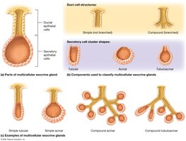
Exocrine Glands
Unicellular Glands: Goblet cells secrete mucus in digestive and respiratory tracts.
Multicellular Glands: Classified by duct structure (simple or compound) and shape (tubular, acinar, tubuloacinar).


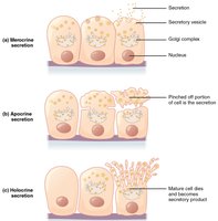
Modes of Exocrine Secretion
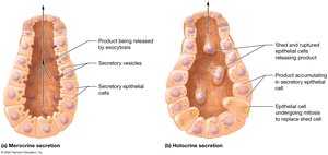
Merocrine: Products released by exocytosis; cell remains intact (e.g., salivary, sweat glands).
Holocrine: Product accumulates, cell ruptures and dies; cell becomes part of secretion (e.g., sebaceous glands).
Apocrine: Apical portion of cell pinched off with product (e.g., mammary glands).

Connective Tissues
Functions and Classification
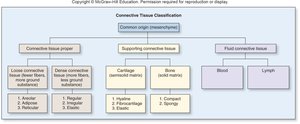
Connective tissues bind, support, protect, and transport substances. They are classified as:
Connective Tissue Proper: Loose, dense, reticular, adipose tissues.
Specialized (Supporting) Connective Tissues: Cartilage, bone, blood.
Cells of Connective Tissue Proper
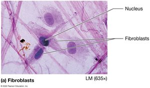
Fibroblasts: Produce protein fibers, ground substance, ECM elements.
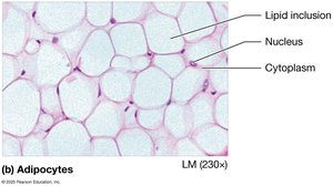
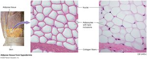
Adipocytes: Fat cells with lipid inclusions.
Mast Cells: Immune cells with granules containing inflammatory mediators.
Phagocytes: Immune cells that engulf foreign substances.
Types of Connective Tissue Proper
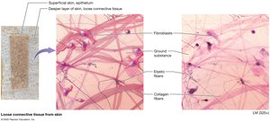
Loose (Areolar) Connective Tissue: Ground substance, all three fiber types, fibroblasts, immune cells; supports blood vessels.
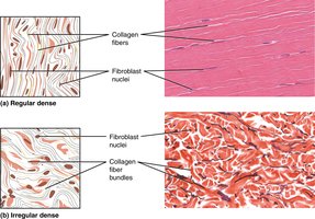
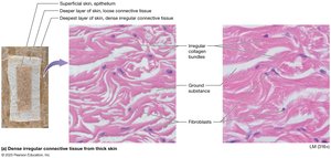
Dense Irregular Connective Tissue: Collagen fibers arranged haphazardly; resists tension in all directions; found in dermis, around organs.
Dense Regular Collagenous Connective Tissue: Parallel collagen fibers; resists tension in one direction; found in tendons, ligaments.
Dense Regular Elastic Connective Tissue: Parallel elastic fibers; allows stretching; found in large blood vessels, some ligaments.
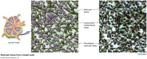
Reticular Tissue: Reticular fibers form networks; support and trap foreign cells; found in lymph nodes.
Adipose Tissue: Fat tissue; insulation, protection, energy reserve; found deep to skin.


Specialized Connective Tissues
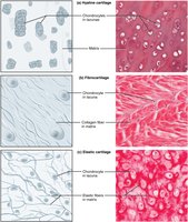
Cartilage
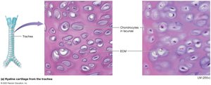
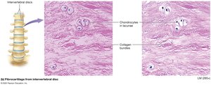
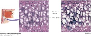
Cartilage is tough, flexible, absorbs shock, and resists tension and compression. It contains chondroblasts (immature) and chondrocytes (mature, in lacunae), and is surrounded by perichondrium.
Hyaline Cartilage: Fine collagen bundles; covers ends of bones, nose, respiratory tract.
Fibrocartilage: Bundles of collagen; found in intervertebral discs, articular discs.
Elastic Cartilage: Elastic fibers; found in external ear, larynx.
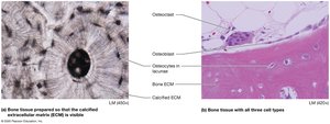
Bone
Bone supports the body, protects organs, stores calcium, and houses bone marrow. Its ECM is 35% organic (collagen, osteoid) and 65% inorganic (calcium phosphate).
Osteoblasts: Bone deposition.
Osteocytes: Maintenance.
Osteoclasts: Bone resorption.
Blood
Blood is a fluid connective tissue with plasma as ECM. Cells include erythrocytes (oxygen transport), leukocytes (immunity), and platelets (clotting).
Muscle Tissues
Types of Muscle Tissue
Muscle cells (myocytes) are excitable and contract to produce movement. Types include:
Skeletal Muscle: Voluntary, multinucleate, attached to skeleton.
Cardiac Muscle: Involuntary, branched, single nucleus, intercalated discs.
Smooth Muscle: Involuntary, flattened, single nucleus, found in hollow organs.
Nervous Tissue
Structure and Function
Nervous tissue makes up the brain, spinal cord, and nerves. It contains neurons (generate, conduct, receive impulses) and neuroglial cells (support, anchor, monitor, speed transmission, circulate fluid).
Neurons: Cell body, axon, dendrites; amitotic.
Neuroglial Cells: Supportive, can divide by mitosis.
Membranes
Types of Membranes
Membranes are thin sheets of tissue lining body surfaces or cavities. Types include:
Serous Membranes: Line body cavities, produce serous fluid.
Synovial Membranes: Line joint cavities, produce synovial fluid.
Mucous Membranes: Line passages opening to outside, secrete mucus.
Cutaneous Membrane: Skin.
Tissue Repair
Regeneration and Fibrosis
Tissue repair is the process of wound healing, which may occur by regeneration (replacement with same cell type) or fibrosis (formation of scar tissue).
Regeneration: Epithelial and most connective tissues.
Fibrosis: Cartilage, cardiac and skeletal muscle, neurons.
Embryonic Origin of Tissues
Germ Layers
Tissues develop from three primary germ layers:
Ectoderm: Forms external tissues, some epithelia.
Mesoderm: Forms mesenchyme, all connective tissues.
Endoderm: Forms internal tissues.
Summary
This guide provides an overview of histology, the structure and function of tissues, their classification, and their roles in the human body. Understanding these concepts is fundamental for further study in anatomy and physiology.
