 Back
BackHistology: Structure and Function of Tissues
Study Guide - Smart Notes
 Tailored notes based on your materials, expanded with key definitions, examples, and context.
Histology: The Study of Tissues
Introduction to Histology
Histology is the branch of anatomy that studies the microscopic structure of tissues. Tissues are groups of cells with similar structure and function, surrounded by an extracellular matrix (ECM) that provides structural and biochemical support.
Types of Tissues
Overview of the Four Primary Tissue Types
Epithelial Tissues (Epithelia): Tightly packed sheets of cells with little to no visible ECM. They cover and line all body surfaces and cavities and form glands that produce secretions or hormones.
Connective Tissues (CT): Characterized by a prominent ECM with scattered cells. They bind, support, protect, and transport substances throughout the body.
Muscle Tissues: Specialized for contraction, enabling movement of the skeleton, heart, and substances through hollow organs.
Nervous Tissues: Composed of neurons (which generate and transmit electrical signals) and neuroglia (which support neuronal function).
The Extracellular Matrix (ECM)
Composition and Function
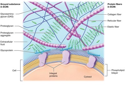
The ECM is a complex network of substances in a liquid, gel, or solid form that surrounds cells. It provides strength, directs cell positioning, and regulates cell development and survival.
Ground Substance: The main component of ECM, consisting of water, nutrients, ions, and macromolecules such as glycosaminoglycans (GAGs), proteoglycans, and cell-adhesion molecules (CAMs).
Protein Fibers: Collagen (resistant to tension), elastic (allow stretch and recoil), and reticular fibers (form supportive meshworks).
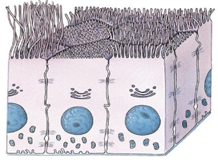
Epithelial Tissues
Functions and Characteristics
Epithelial tissues are found on all internal and external body surfaces. They function in protection, immune defense, secretion, selective transport, and sensation. Epithelia are avascular, consist of tightly packed cells, and rest on a basement membrane (BM).
Classification of Epithelia
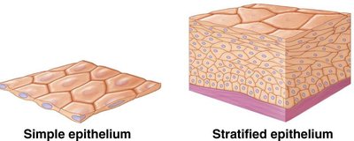
By Layers: Simple (one layer), stratified (multiple layers), and pseudostratified (appears layered but is not).
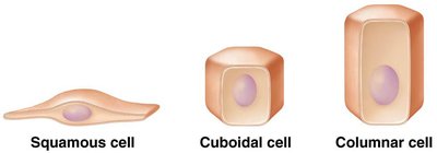
By Cell Shape: Squamous (flat), cuboidal (cube-shaped), columnar (tall and elongated).
Types of Simple Epithelia
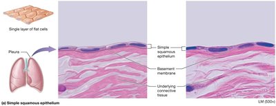
Simple Squamous Epithelium: Thin, single layer for rapid diffusion; found in lung air sacs, kidney, and blood vessel lining.
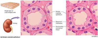
Simple Cuboidal Epithelium: Single layer of cube-shaped cells; found in renal tubules and gland ducts.
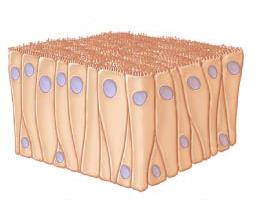
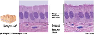
Simple Columnar Epithelium: Single layer of tall cells, often with microvilli or cilia; found in digestive tract lining.
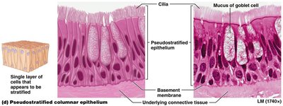
Pseudostratified Ciliated Columnar Epithelium: Appears layered, but all cells touch the basement membrane; found in respiratory tract.
Types of Stratified Epithelia
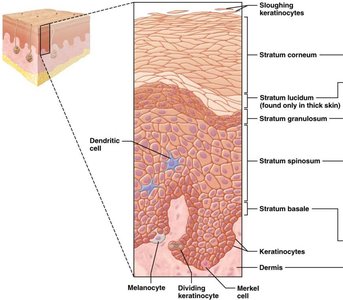
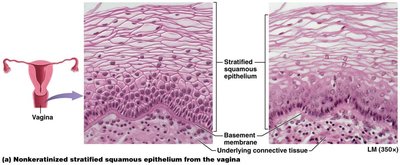
Stratified Squamous Epithelium: Protective; keratinized (skin) or nonkeratinized (mouth, esophagus, vagina).
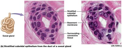
Stratified Cuboidal Epithelium: Rare; lines ducts of sweat glands.
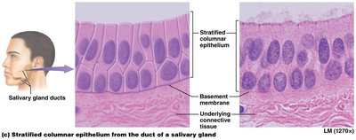
Stratified Columnar Epithelium: Rare; found in male urethra and salivary gland ducts.
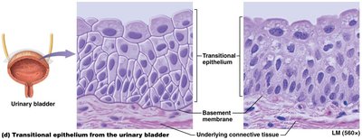
Transitional Epithelium: Found only in the urinary system; cells change shape to allow stretching.
Glandular Epithelia
Types of Glands
Endocrine Glands: Ductless; secrete hormones directly into the bloodstream for systemic effects (e.g., thyroid, pituitary).
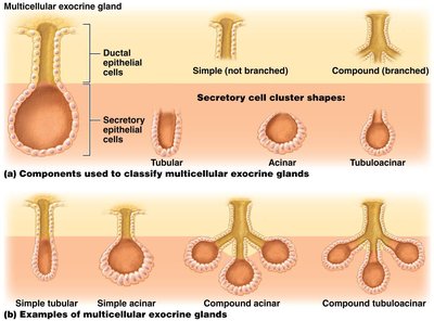
Exocrine Glands: Have ducts; secrete products locally (e.g., sweat, saliva). Can be unicellular (goblet cells) or multicellular.
Modes of Secretion
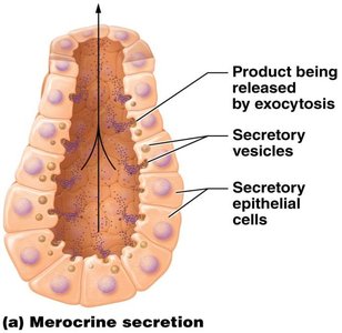
Merocrine Secretion: Products released by exocytosis (e.g., sweat, salivary glands).
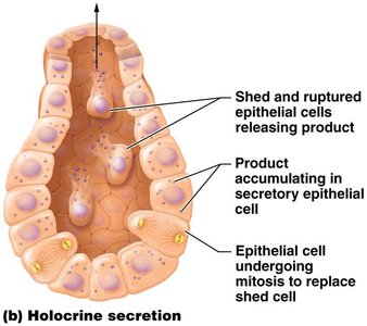
Holocrine Secretion: Entire cells rupture to release products (e.g., sebaceous glands).
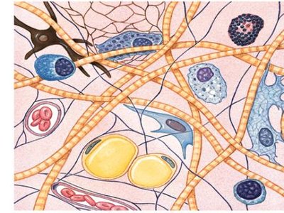
Connective Tissue
Functions and Characteristics
Connective tissues are widely distributed and serve to connect, bind, support, protect, and transport substances. They are characterized by cells embedded in an ECM and are usually vascular.
Types of Connective Tissue Proper
Loose (Areolar) CT: Mostly ground substance, supports epithelia and organs.
Dense CT: Regular (parallel collagen bundles in tendons/ligaments), irregular (disorganized bundles in dermis), and elastic (elastic fibers in large arteries).
Reticular Tissue: Network of reticular fibers supporting lymphoid organs.
Adipose Tissue: Fat storage, insulation, and protection.
Specialized Connective Tissues
Cartilage: Rigid, avascular matrix; types include hyaline (joints, nose), fibrocartilage (intervertebral discs), and elastic (ear).
Bone: Hard matrix, supports and protects, site of blood cell formation.
Blood: Fluid ECM (plasma), transports cells and substances.
Muscle Tissue
Types and Functions
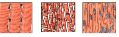
Muscle tissues are specialized for contraction and movement. There are three types:
Skeletal Muscle: Striated, voluntary, attached to bones.
Cardiac Muscle: Striated, involuntary, found in the heart, contains intercalated discs.
Smooth Muscle: Non-striated, involuntary, found in walls of hollow organs and blood vessels.
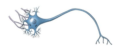
Nervous Tissue
Structure and Function
Nervous tissue is found in the brain, spinal cord, and nerves. It consists of neurons, which transmit electrical signals, and neuroglial cells, which support and protect neurons.
Tissues in Organs and Membranes
Integration of Tissues in Organs
Organs are composed of two or more tissue types that work together for specific functions. For example, skeletal muscle contains both muscle and connective tissue, while the trachea contains multiple tissue layers for air conduction.
Membranes
Serous Membranes: Line body cavities (pericardial, pleural, peritoneal).
Synovial Membranes: Line joint cavities, composed of connective tissue.
Mucous Membranes: Line tubes/organs connecting to the outside (e.g., nasal, oral cavities).
Cutaneous Membrane: The skin.
Tissue Repair
Regeneration and Fibrosis
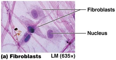
Tissue repair involves regeneration (replacement with the same cell type) or fibrosis (formation of scar tissue by fibroblasts). The process depends on the tissue's regenerative capacity.
Concept Check
Why do the pharynx, esophagus, anus, and vagina have the same organization of epithelium? These areas are subject to wear and tear, requiring protective stratified squamous epithelium.
What is the secretion type where secretory cells rupture to release their contents? Holocrine secretion.
What type of gland releases its product directly into the ECF without ducts? Endocrine gland.
What is the function of an epithelial surface with many microvilli? To increase surface area for absorption.
