 Back
BackHistology Study Guide: Tissue Types, Functions, and Identification
Study Guide - Smart Notes
 Tailored notes based on your materials, expanded with key definitions, examples, and context.
Histology: The Study of Tissues
Overview of Tissue Types
Histology is the study of tissues, which are groups of cells organized to perform specific functions. In animals, there are four principal tissue types: epithelial, connective, muscular, and nervous. Each type has distinct structural and functional characteristics.
Epithelial Tissue: Covers surfaces, lines cavities, and forms glands.
Connective Tissue: Provides support, binds structures, and stores energy.
Muscle Tissue: Facilitates movement through contraction.
Nervous Tissue: Conducts electrical impulses for communication.
Epithelial Tissue
Classification and Functions
Epithelial tissues line body surfaces and cavities, and form glands. They are primarily avascular (lacking blood vessels) and are separated from underlying connective tissue by a basement membrane.
Layering:
Simple Epithelium: One layer; ideal for absorption and secretion (e.g., lungs, digestive tract).
Stratified Epithelium: Multiple layers; protection against abrasion (e.g., skin, esophagus).
Pseudostratified Epithelium: Appears multi-layered but is a single layer of cells of varying heights.
Cell Shape:
Squamous: Flat, scale-like cells.
Cuboidal: Cube-shaped cells.
Columnar: Tall, column-like cells; nuclei are oblong.
Special Types & Surface Modifications:
Transitional Epithelium: Found in the urinary bladder; changes shape as tissue stretches.
Cilia: Hair-like projections for moving substances (e.g., trachea).
Microvilli: Increase surface area for absorption (e.g., small intestine).
Glandular Epithelium:
Exocrine: Uses ducts for secretion (e.g., goblet cells).
Endocrine: Ductless; secretes hormones into the bloodstream (e.g., pancreas).
Connective Tissue (CT)
Classification and Functions
Connective tissues provide internal support and are derived from embryonic mesenchyme. They are mostly composed of an extracellular matrix (fibers and ground substance) and are usually highly vascular.
Matrix Components:
Fibers: Collagen (strength), Elastic (flexibility), Reticular (filtration scaffold).
Ground Substance: Jelly-like material binding cells and fibers.
Loose Connective Tissue:
Areolar: Binds tissue layers, water reservoir.
Adipose: Stores fat, insulation, protection.
Reticular: Scaffold for white blood cells (e.g., spleen).
Dense Connective Tissue:
Dense Regular: Parallel collagen fibers (tendons, ligaments); strong, avascular.
Dense Irregular: Densely-woven network (dermis); omni-directional strength.
Specialized Connective Tissue:
Cartilage: Chondrocytes in lacunae; types include hyaline, fibrocartilage, elastic.
Bone (Osseous): Osteocytes in calcified matrix; compact bone organized into osteons.
Blood: Liquid matrix (plasma) with erythrocytes, leukocytes, thrombocytes.
Muscle and Nervous Tissue
Muscle Tissue
Muscle tissue is contractile, irritable, elastic, and extensible. It is classified as:
Skeletal Muscle: Striated, multinucleate, voluntary control.
Cardiac Muscle: Striated, branched, intercalated discs, involuntary.
Smooth Muscle: Non-striated, uninucleate, found in hollow organs, involuntary.
Nervous Tissue
Nervous tissue consists of neurons (impulse generators) and neuroglia (support cells). Multipolar neurons are most common, with a cell body, many dendrites, and one axon.
Practical Histology: Using the Microscope
Factors Affecting Tissue Appearance
Tissue Source: Slides may contain multiple tissue types.
Sectioning Plane: Tissues can look different when cut transversely or longitudinally.
Stains: Most tissues are transparent and must be dyed. Hematoxylin stains nuclei dark purple; Eosin stains cytoplasm pink/orange.
Identification of Tissue Types (Practice Questions)
Microscopic Images of Tissues
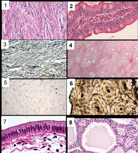
The following images are used for practice in identifying tissue types, their functions, and locations in the body.
Image 1: Dense Regular Connective Tissue (e.g., tendon or ligament). Function: Provides strong attachment; Location: Tendons, ligaments.
Image 2: Simple Columnar Epithelium. Function: Absorption and secretion; Location: Digestive tract lining.
Image 3: Areolar (Loose) Connective Tissue. Function: Binds tissues, supports organs; Location: Under epithelia, around blood vessels.
Image 4: Hyaline Cartilage. Function: Provides support, flexibility; Location: Nose, trachea, ends of long bones.
Image 5: Adipose Tissue. Function: Stores fat, insulation; Location: Under skin, around organs.
Image 6: Compact Bone (Osseous Tissue). Function: Support, protection; Location: Bones of skeleton.
Image 7: Pseudostratified Ciliated Columnar Epithelium. Function: Moves mucus; Location: Trachea, upper respiratory tract.
Image 8: Transitional Epithelium. Function: Allows stretching; Location: Urinary bladder.
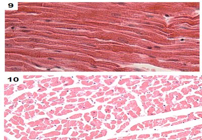
Sectioning Planes in Muscle Tissue
Images 9 and 10 show the same tissue (skeletal muscle) sectioned in two different planes:
Image 9: Longitudinal Section of Skeletal Muscle. Function: Voluntary movement; Location: Attached to bones.
Image 10: Cross Section of Skeletal Muscle. Function: Voluntary movement; Location: Attached to bones.
Sectioning planes affect the appearance of tissues under the microscope. Longitudinal sections show fibers running parallel, while cross sections show circular profiles of fibers.
Blood Supply in Tissues
Vascularity of Tissue Types
Highly Vascular: Areolar, adipose, bone, blood.
Poorly Vascular: Dense regular connective tissue (tendons, ligaments), cartilage.
Avascular: Most epithelial tissues.
Classification of Connective Tissues
Type | Examples | Function | Vascularity |
|---|---|---|---|
Loose | Areolar, Adipose, Reticular | Support, insulation, binding | Good |
Dense | Dense Regular, Dense Irregular | Strength, support | Poor (especially regular) |
Specialized | Cartilage, Bone, Blood | Support, protection, transport | Varies (cartilage poor, bone good, blood excellent) |
Analogy for Tissue Classification
Tissues can be compared to building materials:
Epithelial: Paint or wallpaper—covers surfaces, controls passage.
Connective: Structural frame and insulation—wood, nails, glue.
Muscle: Elevators and plumbing—movement and contraction.
Nervous: Electrical wiring—signal transmission and coordination.
Staining Techniques
Most tissues are transparent and require staining for visualization. The most common stain is Hematoxylin and Eosin (H&E):
Hematoxylin: Stains nuclei dark purple.
Eosin: Stains cytoplasm pink/orange.
Summary Table: Tissue Types, Functions, and Locations
Tissue Type | Function | Location |
|---|---|---|
Simple Columnar Epithelium | Absorption, secretion | Digestive tract |
Pseudostratified Ciliated Columnar Epithelium | Moves mucus | Trachea |
Transitional Epithelium | Stretching | Urinary bladder |
Areolar Connective Tissue | Binding, support | Under epithelia |
Adipose Tissue | Fat storage, insulation | Under skin, around organs |
Dense Regular Connective Tissue | Strong attachment | Tendons, ligaments |
Hyaline Cartilage | Support, flexibility | Nose, trachea, joints |
Compact Bone | Support, protection | Skeletal bones |
Skeletal Muscle | Voluntary movement | Attached to bones |
