 Back
Ch. 4 - Histology: The Study of Tissues
Study Guide - Smart Notes
 Tailored notes based on your materials, expanded with key definitions, examples, and context.
Histology
Introduction to Histology
Histology is the study of tissues, which are groups of cells related in structure and function, surrounded by an extracellular matrix (ECM). Understanding tissues is fundamental to anatomy and physiology, as tissues form the basis of all organs and body systems.
Tissue: A group of similar cells performing a common function.
Extracellular Matrix (ECM): Non-cellular material surrounding cells, providing structural and biochemical support.
Microscopy is essential for studying tissues, as most tissue structures are microscopic.
Types of Tissues
Major Tissue Classes
The human body contains four major tissue types, each defined by the kind and number of cells, the amount and composition of ECM, and their specific functions.
Epithelial tissue
Connective tissue
Muscle tissue
Nervous tissue
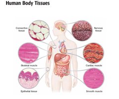
Each tissue type has unique roles in the body, contributing to structure, protection, movement, and communication.
Epithelial Tissue
Characteristics of Epithelial Tissue
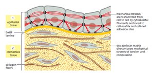
Epithelial tissue forms protective barriers and is specialized for absorption, secretion, and sensation. It covers body surfaces, lines cavities, and forms glands.
Cellularity: Composed almost entirely of tightly packed cells.
Special Contacts: Cells are joined by tight junctions and desmosomes, which determine the tissue's permeability to water and solutes.
Polarity: Has distinct apical (top) and basal (bottom) surfaces.
Supported by Connective Tissue: The basal surface is attached to a basement membrane.
Avascular but Innervated: Lacks blood vessels but contains nerve fibers.
Regenerative: Rapidly replaces lost cells by cell division.
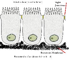
Types of Epithelial Tissue
Simple: One cell layer thick (for absorption, secretion, filtration).
Stratified: Multiple layers (for protection).
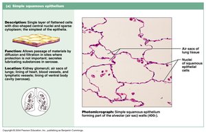
Simple Squamous Epithelium
Single layer of flat cells with disc-shaped nuclei. Functions in diffusion and filtration, providing a slick, friction-reducing lining in lymphatic and cardiovascular systems. Found in lungs, blood vessels, and serous membranes.
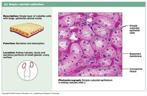
Simple Cuboidal Epithelium
Single layer of cube-like cells with large, central nuclei. Functions in secretion and absorption. Found in kidney tubules, ducts of glands, and ovary surface.
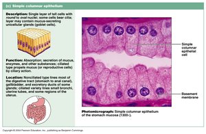
Simple Columnar Epithelium
Single layer of tall cells, often with cilia or microvilli. Functions in absorption and secretion; may contain goblet cells. Found in the digestive tract, uterine tubes, and some regions of the uterus.
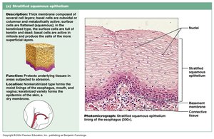
Stratified Squamous Epithelium
Thick membrane composed of several layers. Functions in protection against abrasion. Forms the external part of the skin (keratinized) and linings of the esophagus, mouth, and vagina (nonkeratinized).
Connective Tissue
Structural Elements of Connective Tissue
Connective tissue supports, binds, and protects other tissues and organs. It consists of cells, fibers, and ground substance (ECM).
Ground Substance: Unstructured material filling the space between cells.
Fibers: Collagen (strength), elastic (stretch), reticular (supportive networks).
Cells: Fibroblasts (produce fibers), chondroblasts (cartilage), osteoblasts (bone), hematopoietic stem cells (blood), and others.
Types of Connective Tissue Proper
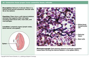
Reticular Connective Tissue
Loose ground substance with reticular fibers; forms a soft internal skeleton (stroma) supporting other cell types. Found in lymph nodes, bone marrow, and spleen.
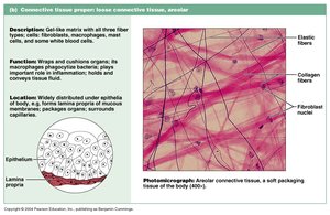
Areolar Connective Tissue
Gel-like matrix with all three fiber types. Wraps and cushions organs; widely distributed under epithelia.
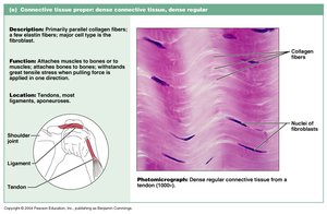
Dense Regular Connective Tissue
Parallel collagen fibers with few elastic fibers. Attaches muscles to bones or to other muscles, and bone to bone. Found in tendons, ligaments, and aponeuroses.
Dense Irregular Connective Tissue
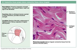
Irregularly arranged collagen fibers with some elastic fibers. Withstands tension in many directions; found in the dermis, submucosa of the digestive tract, and fibrous organ capsules.
Cartilage
Hyaline Cartilage
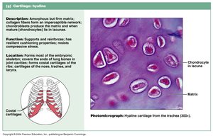
Firm matrix with collagen fibers; chondrocytes in lacunae. Supports, reinforces, cushions, and resists compression. Found in embryonic skeleton, ends of long bones, nose, trachea, and larynx.
Fibrocartilage
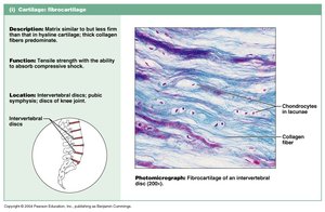
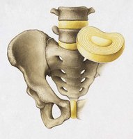
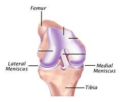
Matrix similar to hyaline but less firm, with thick collagen fibers. Provides tensile strength and absorbs compression shock. Found in intervertebral discs, pubic symphysis, and knee menisci.
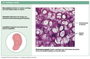
Elastic Cartilage
Similar to hyaline cartilage but with more elastic fibers. Maintains shape and structure while allowing flexibility. Supports external ear (pinna) and epiglottis.
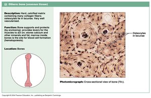
Bone (Osseous Tissue)
Hard, calcified matrix with collagen fibers; osteocytes in lacunae. Well vascularized. Stores calcium, minerals, and fat; marrow is the site of hematopoiesis (blood cell formation).
Glands
Classification of Glands
A gland is one or more cells that makes and secretes an aqueous fluid. Glands are classified by their site of release and the number of cells forming the gland.
Endocrine glands: Ductless; release hormones into the blood or interstitial fluid.
Exocrine glands: Secrete products onto body surfaces or into body cavities; may be unicellular (e.g., goblet cells) or multicellular (e.g., sweat glands).
Exocrine glands can be further classified by the type of secretion: serous (watery), mucus (mucin-rich), or mixed.
Nervous Tissue
Structure and Function
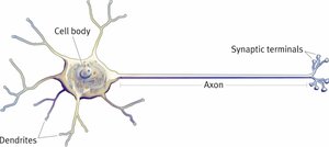
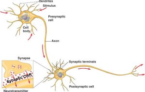
Nervous tissue consists of nerve cells (neurons) and supporting cells. Neurons are specialized for communication, with three main parts: dendrites (receive information), cell body (processes information), and axon/axon terminals (transmit signals).
Synapse
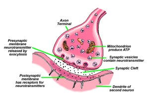
A synapse is a close connection between a neuron and another cell (neuron, muscle, or gland), allowing for the transmission of signals via neurotransmitters.
Muscle Tissue
Types of Muscle Tissue
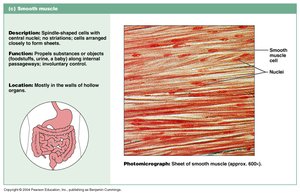
Muscle tissue is specialized for contraction and movement. There are three types:
Skeletal muscle: Voluntary movement, attached to bones.
Cardiac muscle: Involuntary, found in the heart.
Smooth muscle: Involuntary, found in walls of hollow organs.
Skeletal Muscle Structure
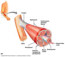
Skeletal muscle is organized into bundles of muscle fibers, which are grouped into larger bundles and surrounded by connective tissue coverings. This organization allows for efficient force generation and transmission.
Additional info: The study of tissues (histology) is foundational for understanding organ structure and function in anatomy and physiology. Knowledge of tissue types and their properties is essential for interpreting pathological changes in disease.
