 Back
BackHistology: The Study of Tissues – Epithelial and Connective Tissue Overview
Study Guide - Smart Notes
 Tailored notes based on your materials, expanded with key definitions, examples, and context.
Histology: The Study of Tissues
Introduction to Histology
Histology is the microscopic study of tissues, which are groups of cells that work together to perform specific functions. The human body is composed of four basic tissue types, each with unique structures and functions.
Epithelial tissue
Connective tissue
Muscle tissue
Nervous tissue
All tissues consist of specialized cells and an extracellular matrix (ECM), which is produced by the tissue's cells.
Epithelial Tissue
General Features of Epithelial Tissue
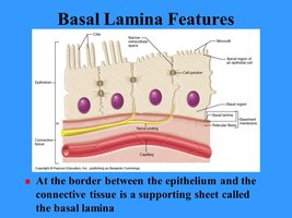
Epithelial tissue covers body surfaces, lines body passageways and cavities, and forms glands. Its ECM is mostly limited to the basal lamina, a thin layer beneath the cells. The basal lamina adheres to the lamina reticularis, another ECM layer produced by underlying connective tissue. Together, these layers form the basement membrane, which supports and anchors the epithelium.
Vascularity and Thickness
Epithelial tissues are avascular (lack blood vessels) and rely on diffusion from underlying tissues for nutrients and oxygen. This limits their thickness, as cells too far from the blood supply cannot survive.
Classification of Epithelial Tissue
Epithelia are classified by two main criteria:
Number of cell layers (simple vs. stratified)
Shape of the cells (squamous, cuboidal, columnar)
Simple Epithelia
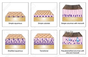
Simple epithelia have a single layer of cells and are specialized for absorption, secretion, and filtration.
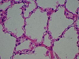
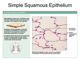
Simple squamous epithelium: Single layer of flat cells with flattened nuclei. Found in alveoli of lungs, blood vessels, and kidney glomeruli. Allows rapid diffusion and filtration.
Simple cuboidal epithelium: Single layer of cube-shaped cells with round, central nuclei. Found in glands (thyroid, salivary, mammary), kidney tubules, and some respiratory passages. Functions in absorption and secretion.
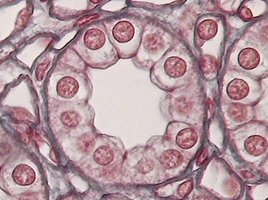
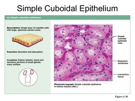
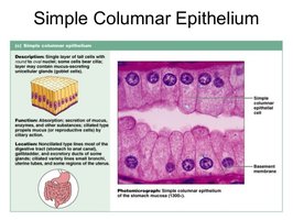
Simple columnar epithelium: Single layer of tall, column-shaped cells with nuclei near the base. Found in digestive tract, gall bladder, uterine tubes, and some kidney tubules. May have cilia or microvilli; often contains goblet cells that secrete mucus.
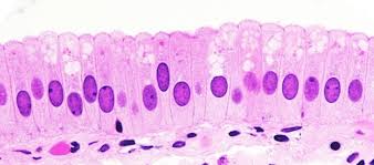
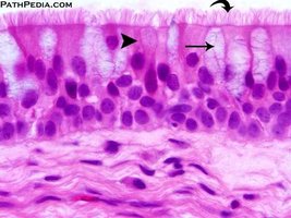
Pseudostratified Ciliated Columnar Epithelium
This tissue appears to have multiple layers due to varying cell heights, but all cells rest on the basement membrane (pseudo = false). Usually ciliated and found in the respiratory tract and nasal cavity.
Stratified Epithelia
Stratified epithelia have two or more layers of cells and are specialized for protection against abrasion and mechanical stress.
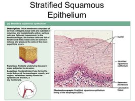
Stratified squamous epithelium: Multiple layers of flat cells. Two main types:
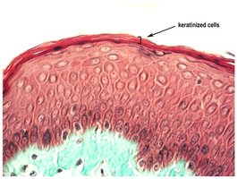
Keratinized: Surface cells are dead and filled with keratin, providing a tough, waterproof barrier. Found in the epidermis of the skin.
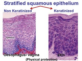
Non-keratinized: Surface cells are alive, moist, and lack keratin. Found in the oral cavity, pharynx, anus, and vagina.
Other Stratified Epithelia
Stratified cuboidal and stratified columnar epithelium: Rare in the human body; found lining ducts of certain glands.
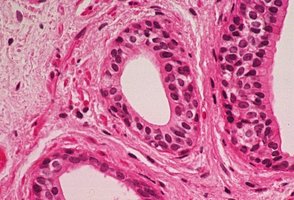
Transitional Epithelium
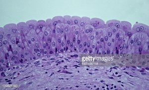
Transitional epithelium is a stratified tissue with cells that can change shape from dome-shaped to squamous when stretched. Found lining the urinary bladder and ureters, allowing these organs to expand and contract.
Connective Tissue
General Features of Connective Tissue
Connective tissue serves to connect, support, and protect other tissues and organs. It contains relatively few cells and a prominent ECM composed of ground substance and protein fibers.
Collagen fibers: Provide strength
Elastic fibers: Provide stretch and recoil
Reticular fibers: Form supportive networks
Types of Connective Tissue
There are four general types of connective tissue:
Connective tissue proper
Cartilage
Bone
Blood
Connective Tissue Proper
Areolar connective tissue: Loose arrangement of fibers and cells, supports and cushions organs, found beneath epithelia and around blood vessels.
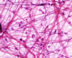
Reticular connective tissue: Network of reticular fibers, forms the stroma of lymphoid organs (spleen, lymph nodes).

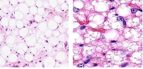
Adipose tissue: Composed of adipocytes that store fat, provides insulation and energy storage, found under skin and around organs.
Dense regular connective tissue: Parallel bundles of collagen fibers, very strong, found in tendons and ligaments.

Dense irregular connective tissue: Collagen fibers arranged irregularly, provides strength in multiple directions, found in dermis and organ capsules.
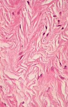
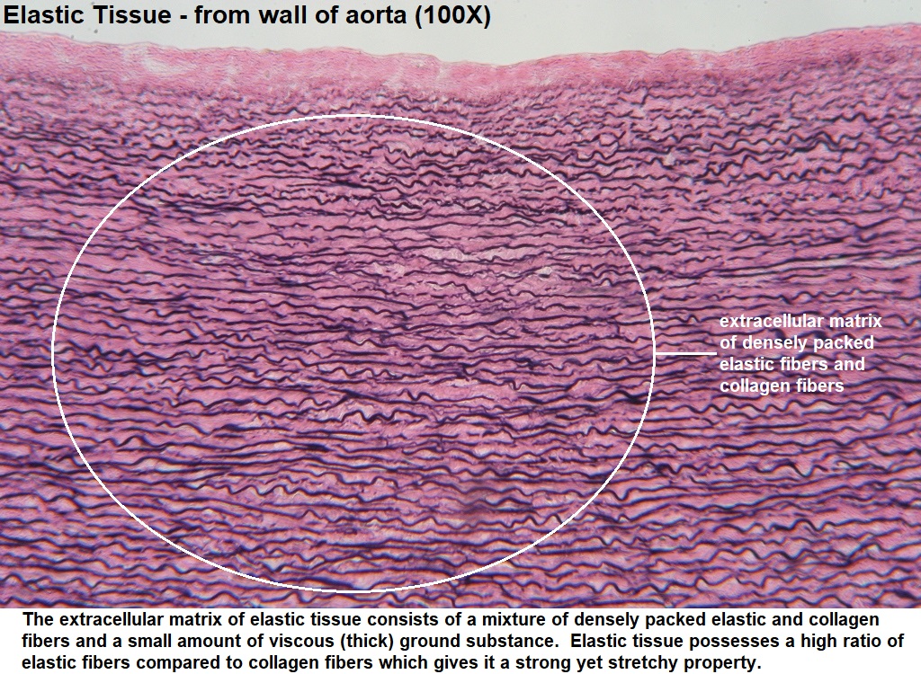
Dense elastic connective tissue: Elastic fibers arranged in parallel, found in large blood vessels and some ligaments.

Cartilage
Cartilage is tough but flexible, resists tension and compression. It contains chondrocytes in lacunae and is avascular. Three types:
Hyaline cartilage: Smooth, glassy appearance, found at ends of bones, nose, and respiratory passages.

Fibrocartilage: Abundant collagen fibers, very strong, found in intervertebral discs and menisci.

Elastic cartilage: Contains elastic fibers, flexible, found in ear and epiglottis.


Bone (Osseous Tissue)
Bone tissue contains osteocytes in a mineralized ECM with collagen and calcium salts. The ECM is arranged in concentric layers (lamellae), making bone the hardest tissue in the body.

Blood
Blood is a connective tissue with a liquid ECM (plasma) and formed elements (erythrocytes, leukocytes, platelets). It functions in transport, immunity, and homeostasis.

Muscle and Nervous Tissue (Overview)
Muscle Tissue
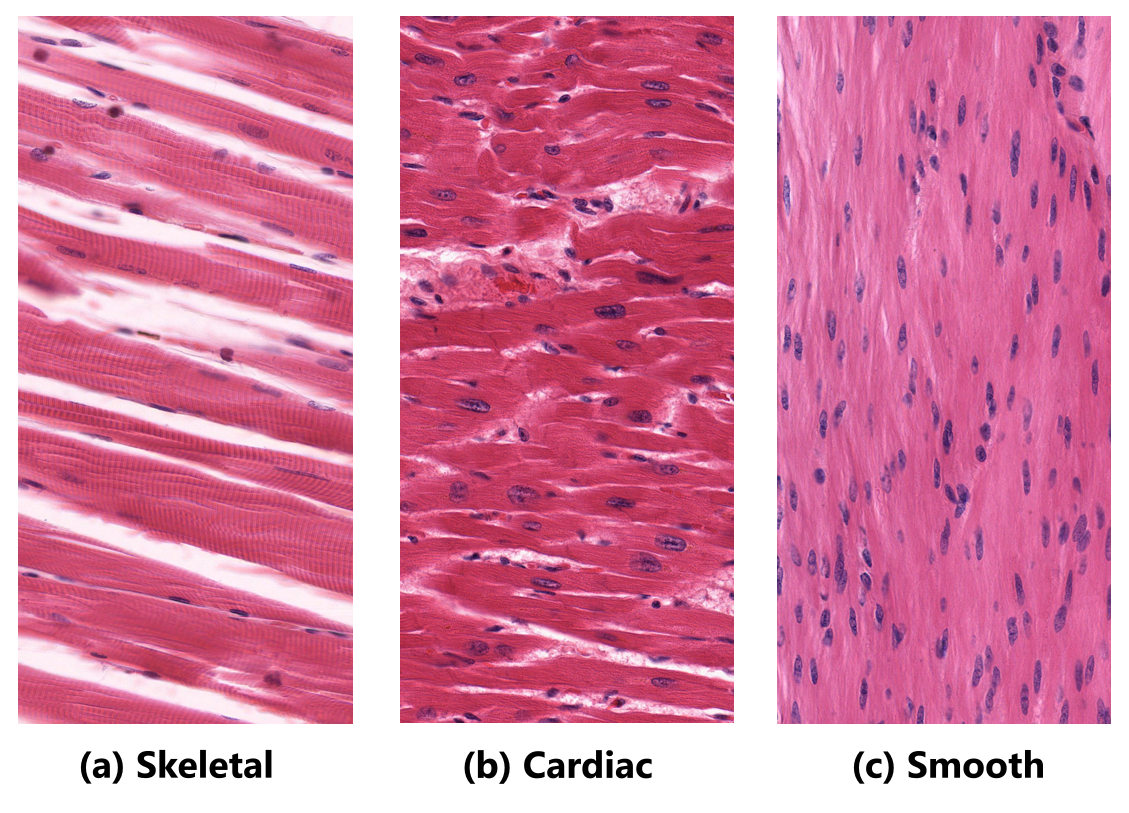
Muscle tissue is specialized for contraction and movement. Types include:
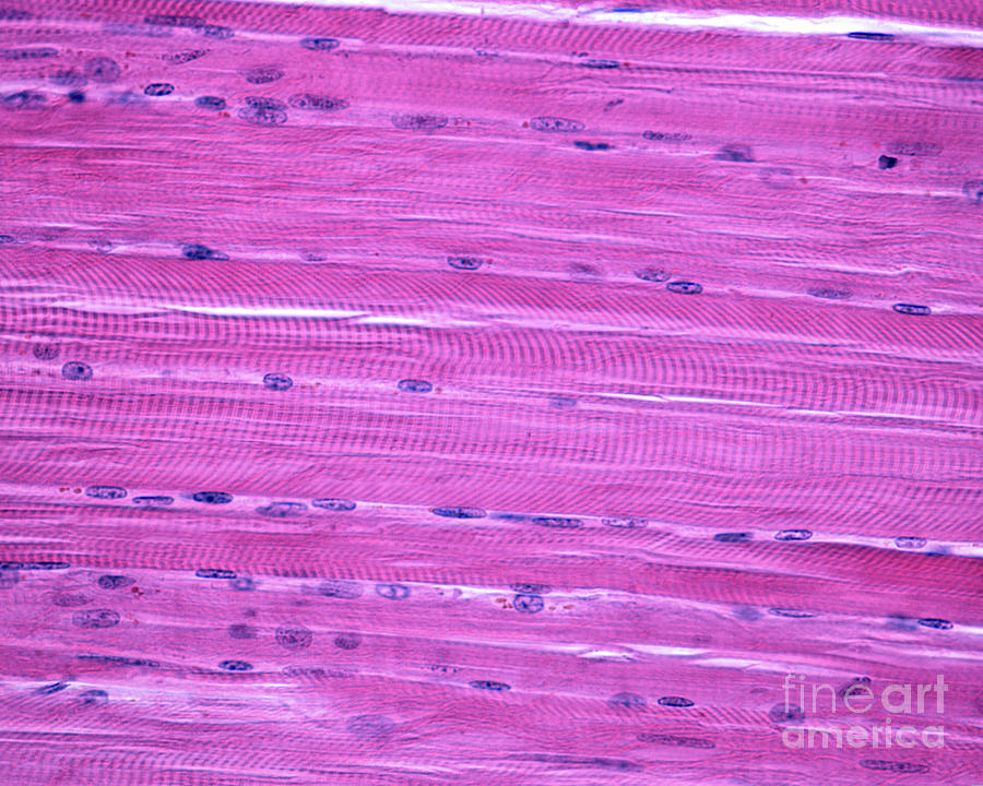
Skeletal muscle: Long, striated, multinucleated fibers, voluntary movement.
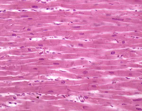
Cardiac muscle: Striated, branched cells with intercalated discs, found in the heart.
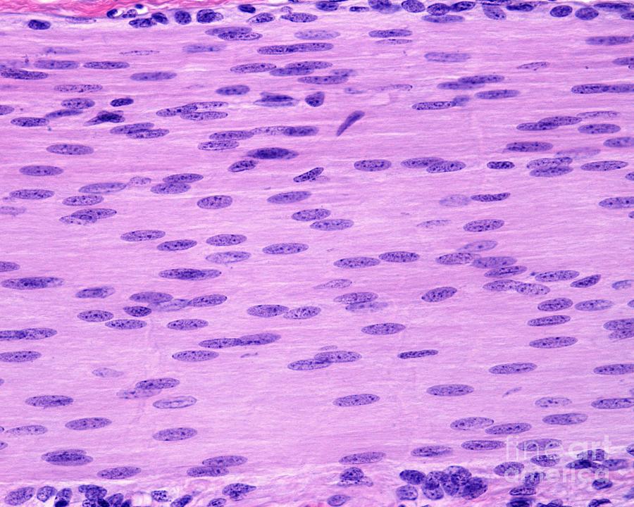
Smooth muscle: Non-striated, spindle-shaped cells, found in walls of hollow organs.
Nervous Tissue
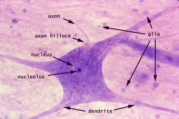
Nervous tissue consists of neurons (cells that transmit electrical signals) and neuroglial cells (supporting cells). Neurons have a cell body, dendrites, and an axon.
Additional info: This guide provides a comprehensive overview of histology, focusing on epithelial and connective tissues, with brief context on muscle and nervous tissues for completeness.
