 Back
BackHistology: The Study of Tissues – Structured Study Notes for Anatomy & Physiology
Study Guide - Smart Notes
 Tailored notes based on your materials, expanded with key definitions, examples, and context.
Histology: The Study of Tissues
Introduction to Tissues & Histology
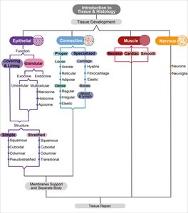
Histology is the scientific study of tissues, focusing on their structure, function, and arrangement. The human body is composed of four primary tissue types: epithelial, connective, muscle, and nervous tissue. Each tissue type plays a distinct role in maintaining the body's structure and function.
Tissue: A group of similar cells working together to perform related functions, often including the extracellular matrix (ECM), which is the material surrounding cells.
Histology: The study of tissue structure and function, including cell shape, size, arrangement, and ECM.
ECM: Complex molecules outside cells, especially prominent in connective tissue.
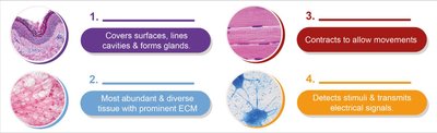
Additional info: Tissues are transparent under a microscope before being stained with dyes for visualization.

Overview of Tissue Types
Epithelial Tissue: Covers surfaces, lines cavities, and forms glands.
Connective Tissue: Most abundant and diverse, with prominent ECM.
Muscle Tissue: Contracts to allow movement.
Nervous Tissue: Detects stimuli and transmits electrical signals.
Cell Junctions
Types of Cell Junctions
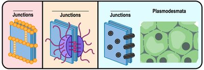
Cell junctions are specialized structures that connect adjacent cells, allowing communication and maintaining tissue integrity. There are four main types:
Tight Junctions: Membrane proteins join neighboring cells, creating an impermeable barrier.
Desmosomes (Anchoring Junctions): Intermediate filaments anchor cells together, providing mechanical strength.
Gap Junctions: Protein channels connect the cytoplasm of two cells, allowing exchange of small molecules and ions.
Plasmodesmata: Gap junctions in plant cells, connecting cytoplasm across cell walls.
Junction Type | Description |
|---|---|
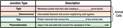
Tight | Membrane protein that links cells creating a waterproof barrier. |
Desmosomes | Intermediate filaments that anchor neighboring cells together. |
Gap | Protein channels that connect the cytoplasm of two animal cells. |
Plasmodesmata | Gaps in the cell walls that connect the cytoplasms of two plant cells. |
Epithelial Tissue
Functions and Locations
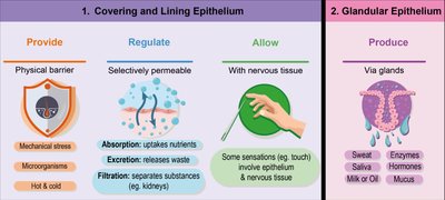
Epithelial tissue forms sheets of tightly packed cells on a basement membrane adjacent to connective tissue. It covers body surfaces, lines internal cavities and ducts, and forms glands.
Protection: Acts as a physical barrier against mechanical stress, microorganisms, and temperature extremes.
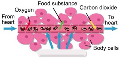
Regulation: Selectively permeable, allowing absorption, excretion, and filtration.
Sensation: Works with nervous tissue to allow sensations such as touch.
Secretion: Produces substances via glands (e.g., sweat, saliva, hormones, mucus).
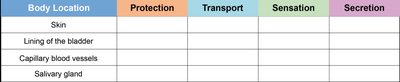
Body Location | Protection | Transport | Sensation | Secretion |
|---|---|---|---|---|
Skin | X | X | ||
Lining of the bladder | X | X | ||
Capillary blood vessels | X | |||
Salivary gland | X |
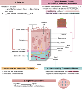
Characteristics of Epithelial Tissue
Polarity: Has distinct apical (top) and basal (bottom) surfaces.
Tightly Pressed Tissue: Anchored to basement membrane, supported by connective tissue.
Avascular but Innervated: No blood vessels, but has nerve supply.
Highly Regenerative: Cells divide regularly, making epithelium prone to cancer.
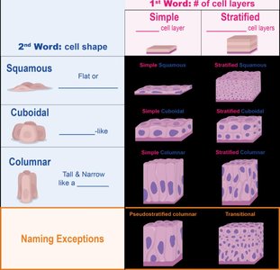
Structural Naming of Epithelial Tissue
Epithelial tissues are named based on the number of cell layers and cell shape:
Simple: One layer of cells.
Stratified: Multiple layers of cells.
Squamous: Flat cells.
Cuboidal: Cube-like cells.
Columnar: Tall and narrow cells.
Types of Simple Epithelial Tissue
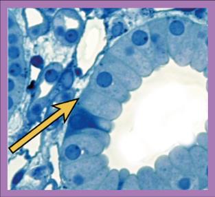
Simple Squamous Epithelium: Single layer of flat cells; ideal for rapid diffusion and lining body cavities.
Simple Cuboidal Epithelium: Single layer of cube-shaped cells; functions in absorption and secretion, found in kidney tubules and ducts.
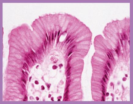
Simple Columnar Epithelium: Single layer of tall, narrow cells; functions in absorption and secretion, found in digestive tract.
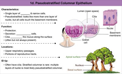
Pseudostratified Columnar Epithelium: Appears layered but all cells touch the basement membrane; found in upper respiratory passages.
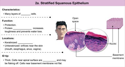
Types of Stratified Epithelial Tissue
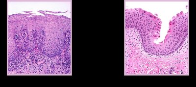
Stratified Squamous Epithelium: Many layers of flat cells; provides protection, found in skin and orifices.
Transitional Epithelium: Many layers, transitions from cuboidal to squamous; found in bladder, ureters, and urethra.
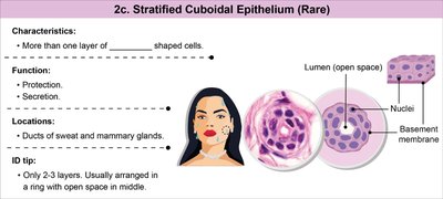
Stratified Cuboidal Epithelium (Rare): More than one layer of cube-shaped cells; found in ducts of sweat and mammary glands.
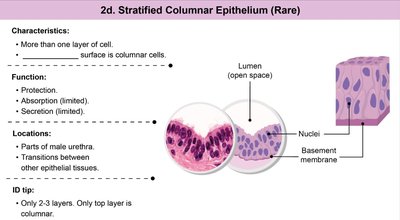
Stratified Columnar Epithelium (Rare): More than one layer, top layer is columnar; found in parts of male urethra.
Glandular Epithelial Tissue
Types of Glands
Exocrine Glands: Release products onto body surfaces or into ducts (e.g., sweat, saliva).
Endocrine Glands: Release products (hormones) directly into the bloodstream.
Glands can be unicellular (e.g., goblet cells) or multicellular, and are classified by their mode of secretion:
Merocrine: Secrete products via exocytosis (most common).
Holocrine: Whole cell ruptures to release product.
Apocrine: Apical portion of cell sheds off, cell survives.
Connective Tissue
Introduction and Structure
Connective tissue is the most abundant and diverse tissue class, characterized by a prominent ECM. All connective tissues are derived from embryonic mesenchyme, and their cells occupy less space than the ECM.
Ground Substance: Unstructured material between cells and fibers, ranges from solid to liquid.
Protein Fibers: Provide strength, flexibility, and recoil.
Cells: "-blast" cells are immature and secrete ECM; "-cyte" cells are mature and maintain ECM.
Classes of Connective Tissue
Connective Tissue Proper: Includes loose and dense connective tissues.
Specialized Connective Tissue: Includes cartilage, bone, blood, and lymph.
Connective Tissue Proper
Loose Connective Tissue: Loosely arranged fibers, often vascular; includes areolar, reticular, and adipose tissue.
Dense Connective Tissue: Densely packed fibers, less vascular; includes regular, irregular, and elastic tissue.
Protein fibers in connective tissue proper include:
Collagen Fibers: Long, thick, straight, strong but flexible.
Reticular Fibers: Thin, branched, netlike, resists force in multiple directions.
Elastic Fibers: Branched, wavy, allows recoil.
Specialized Connective Tissue
Cartilage
Hyaline Cartilage: Smooth appearance, reduces friction.
Fibrocartilage: Large collagen bundles, resists compression and allows flexibility.
Elastic Cartilage: Flexible, found in ear and epiglottis.
Bone
Osteoblasts: Build bone matrix.
Osteocytes: Maintain bone matrix.
ECM: Rigid due to mineralization.
Blood
Fluid matrix: Aids in transport of gases and nutrients.
Red blood cells: Carry oxygen; abnormal shapes can affect viscosity and transport.
Muscle Tissue
Types of Muscle Tissue
Skeletal Muscle: Voluntary, striated, long cylindrical cells, multinucleated.
Cardiac Muscle: Involuntary, striated, branching cells, uninucleate, intercalated discs for coordination.
Smooth Muscle: Involuntary, non-striated, spindle-shaped cells, uninucleate, found in walls of hollow organs.
Nervous Tissue
Structure and Function
Nervous tissue is specialized to generate and carry electrical impulses throughout the body. It is located in the brain, spinal cord, and nerves.
Neurons: Conduct electrical impulses; composed of dendrites (receive signals), cell body (soma), and axon (transmits signals).
Neuroglia (Glial Cells): Support, insulate, and protect neurons; do not conduct impulses.
Serous Membranes
Organization and Function
Serous membranes are thin sheets of tissue forming double-layered membranes that wrap many organs (viscera). They consist of a visceral layer (attached to organs) and a parietal layer (attached to body walls), with a serous cavity between them containing lubricating serous fluid.
Pleurae: Surround each lung.
Pericardium: Surrounds the heart.
Peritoneum: Surrounds most abdominal organs.
Additional info: Serous fluid lubricates membranes, allowing organs to move easily. Inflammation (e.g., pericarditis, pleurisy) can cause pain and affect organ function.
