 Back
BackHistology: The Tissue Level of Organization
Study Guide - Smart Notes
 Tailored notes based on your materials, expanded with key definitions, examples, and context.
Introduction to Histology
Definition and Importance of Tissues
Histology is the study of tissues, which are collections of cells and their products (extracellular matrix) that perform specific, limited functions. The human body is composed of four basic tissue types: epithelial, connective, muscle, and nervous tissue. Each type plays a distinct role in the structure and function of organs and systems.
Epithelial tissue: Covers surfaces, lines cavities, and forms glands.
Connective tissue: Supports, binds, and protects organs; stores energy; provides immunity.
Muscle tissue: Responsible for movement and force generation.
Nervous tissue: Initiates and transmits nerve impulses for coordination.
Cell Junctions
Types and Functions of Cell Junctions
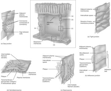
Cell junctions are specialized structures that connect adjacent cells, providing communication, adhesion, and barrier functions. They are essential for maintaining tissue integrity and function.
Tight junctions: Form watertight seals between cells, preventing leakage of fluids (e.g., in the gastrointestinal tract and bladder).
Gap junctions: Allow direct communication between cells via connexons, facilitating the passage of ions and small molecules (e.g., in cardiac and smooth muscle).
Desmosomes: Provide strong adhesion between cells, resisting mechanical stress (e.g., in skin and heart tissue).
Hemidesmosomes: Anchor cells to the basement membrane, stabilizing epithelial layers.
Epithelial Tissue
General Features
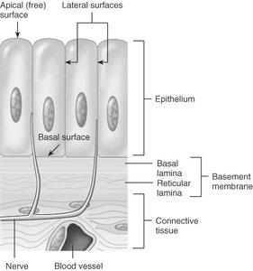
Epithelial tissue consists of closely packed cells with minimal extracellular material. It is avascular (lacks blood vessels), relies on diffusion for nutrient and waste exchange, and exhibits rapid cell division. Epithelial cells are anchored to a basement membrane, which provides support and attachment.
Apical surface: Exposed to the body surface or cavity.
Basal surface: Attached to the basement membrane.
Functions: Protection, absorption, secretion, filtration, excretion, sensory reception, and reproduction.
Classification of Epithelia
Epithelia are classified by the number of cell layers and the shape of the surface cells:
Layers:
Simple: One layer
Stratified: Two or more layers
Pseudostratified: Appears multilayered but all cells contact the basement membrane
Shapes:
Squamous: Flat
Cuboidal: Cube-shaped
Columnar: Tall and column-like
Transitional: Variable shape, stretches
Types of Epithelia and Examples
Simple Squamous Epithelium: Single layer of flat cells; found in lungs, kidneys, blood vessels (endothelium), and body cavities (mesothelium).
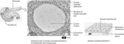
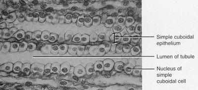
Simple Cuboidal Epithelium: Single layer of cube-shaped cells; lines kidney tubules and ducts, adapted for absorption and secretion.
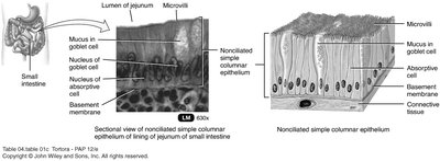
Simple Columnar Epithelium: Single layer of tall cells; may have microvilli (for absorption in GI tract) or cilia (for movement in respiratory tract and uterine tubes).
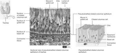
Pseudostratified Columnar Epithelium: Appears multilayered; all cells touch the basement membrane but not all reach the surface; found in respiratory tract, male urethra.
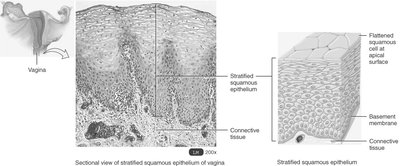
Stratified Squamous Epithelium: Multiple layers; surface cells are flat. Keratinized (skin) or nonkeratinized (mouth, vagina).
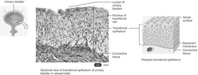
Transitional Epithelium: Multiple layers; surface cells change shape with stretching; lines urinary bladder, ureters, and part of urethra.
Glandular Epithelium
Glandular epithelium forms glands that secrete substances. Glands are classified as exocrine (secrete onto surfaces via ducts) or endocrine (secrete hormones into the bloodstream, ductless).
Exocrine glands: Sweat, salivary, digestive glands; may be unicellular (goblet cells) or multicellular.
Endocrine glands: Thyroid, adrenal, pituitary glands; secrete hormones for homeostasis.
Structural and Functional Classification of Exocrine Glands
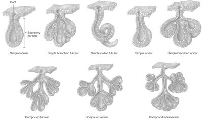
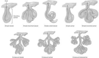
Structural:
Simple (unbranched duct) or compound (branched duct)
Tubular (tube-like) or acinar (flask-like)
Functional:
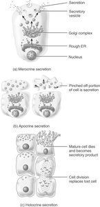
Merocrine: Secrete by exocytosis (e.g., sweat, saliva)
Apocrine: Apical portion pinches off (e.g., some sweat glands)
Holocrine: Entire cell ruptures to release product (e.g., sebaceous glands)
Connective Tissue
General Features
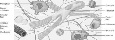
Connective tissue is the most abundant and widely distributed tissue type. It consists of cells and an extracellular matrix (ground substance and fibers). The matrix determines the tissue's properties and can be liquid, gel-like, or solid.
Cells: Fibroblasts, adipocytes, macrophages, plasma cells, mast cells, leukocytes.
Fibers: Collagen (strength), elastic (stretch), reticular (support).
Ground substance: Glycosaminoglycans (GAGs), hyaluronic acid, chondroitin sulfate, adhesion proteins.
Types of Connective Tissue
Embryonic connective tissue: Mesenchyme (gives rise to all other connective tissues), mucous connective tissue (Wharton's jelly in umbilical cord).
Mature connective tissue:
Connective tissue proper: Loose (areolar, adipose, reticular) and dense (regular, irregular, elastic)
Cartilage: Hyaline, fibrocartilage, elastic
Bone: Compact and spongy
Blood and lymph
Loose Connective Tissue
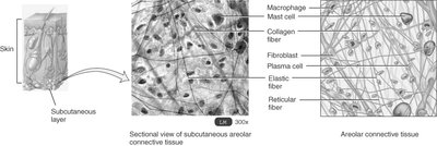
Areolar: All three fiber types, various cells; found in subcutaneous layer.
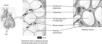
Adipose: Adipocytes store fat; insulates, protects, and stores energy.
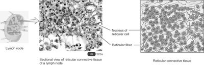
Reticular: Network of reticular fibers; forms stroma of organs like spleen and lymph nodes.
Dense Connective Tissue
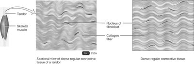
Dense regular: Parallel collagen fibers; forms tendons and ligaments.
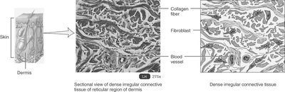
Dense irregular: Irregularly arranged collagen fibers; found in dermis, heart valves.
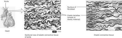
Elastic: Abundant elastic fibers; found in lungs, arteries, vocal cords.
Cartilage
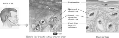
Cartilage is a resilient, avascular connective tissue with chondrocytes in lacunae and a matrix rich in collagen and chondroitin sulfate. Types include:
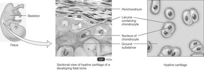
Hyaline cartilage: Most common, flexible, reduces friction at joints.
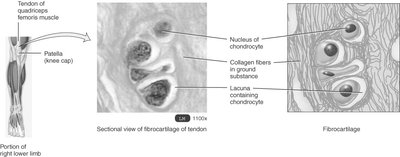
Fibrocartilage: Strongest, resists compression; found in intervertebral discs.
Elastic cartilage: Flexible, maintains shape; found in ear and epiglottis.
Bone (Osseous) Tissue
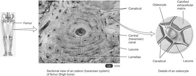
Bone tissue provides support, protection, movement, mineral storage, and blood cell formation. It consists of a mineralized matrix and osteocytes in lacunae. Two types:
Compact bone: Organized into osteons (Haversian systems).
Spongy bone: Contains trabeculae and red marrow.
Blood and Lymph
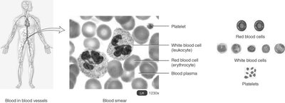
Blood: Liquid matrix (plasma) with red cells (erythrocytes), white cells (leukocytes), and platelets; transports gases, nutrients, wastes, and immune cells.
Lymph: Interstitial fluid in lymphatic vessels; involved in immune response and transport of substances.
Membranes
Types of Membranes
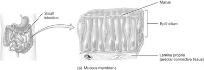
Mucous membranes (mucosae): Line cavities opening to the exterior; secrete mucus for protection and moisture.
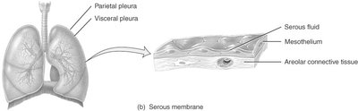
Serous membranes: Line closed body cavities; secrete serous fluid to reduce friction (e.g., pleura, peritoneum, pericardium).
Cutaneous membrane: The skin; covers the body surface.
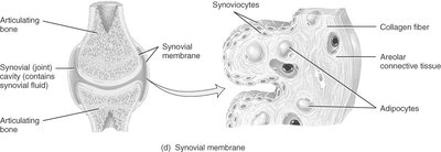
Synovial membranes: Line joint cavities; secrete synovial fluid for lubrication.
Muscle Tissue
Types and Functions
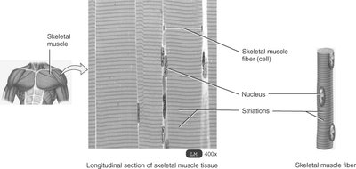
Skeletal muscle: Long, cylindrical, multinucleated, striated; voluntary movement.
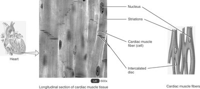
Cardiac muscle: Branched, striated, single nucleus, intercalated discs; involuntary, found in heart wall.
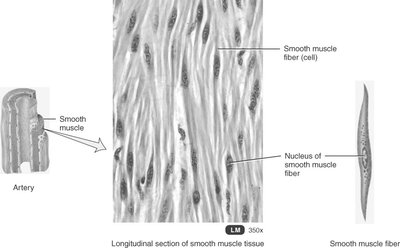
Smooth muscle: Spindle-shaped, nonstriated, single nucleus; involuntary, found in walls of hollow organs.
Nervous Tissue
Structure and Function
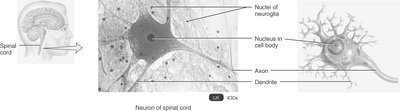
Nervous tissue consists of neurons and neuroglia. Neurons have a cell body, dendrites (receive signals), and an axon (transmits signals). Neuroglia support and protect neurons. Nervous tissue is specialized for communication and control of body functions.
Tissue Repair and Aging
Repair Mechanisms
Epithelial tissue: High capacity for renewal via stem cells.
Connective tissue: Bone repairs well; cartilage repairs slowly.
Muscle tissue: Limited repair capacity.
Nervous tissue: Poorest repair capacity.
Factors Affecting Repair
Nutrition (protein, vitamin C)
Blood circulation
Aging (decreased cell division, altered collagen and elastin)
Fibrosis is scar formation; adhesions are abnormal tissue connections after injury or surgery.
