 Back
Histology: Tissues and the Integumentary System
Study Guide - Smart Notes
 Tailored notes based on your materials, expanded with key definitions, examples, and context.
Histology: The Study of Tissues
Introduction to Tissues
Histology is the study of tissues, which are groups of cells that work together to perform specific functions. The four primary tissue types in the human body are epithelial, connective, muscle, and nervous tissues. Each type has specialized roles essential for the structure and function of organs and systems.
Epithelial Tissue
Characteristics and Functions
Epithelial tissue is specialized for lining and covering body surfaces and cavities, as well as forming glands. It provides protection, immune defense, secretion, regulation of transport, and sensation.
Protection: Shields underlying tissues from mechanical and thermal injury.
Immune Defense: Acts as a barrier against microorganisms.
Secretion: Forms glands that produce substances like oil and hormones.
Transport Regulation: Selectively permeable membranes control molecule passage.
Sensation: Contains nerves to detect environmental changes.
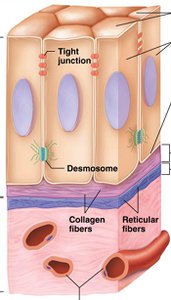
Cell Junctions in Epithelial Tissue
Cell junctions are specialized structures that connect epithelial cells, providing structural integrity and communication.
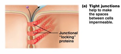
Tight Junctions: Seal spaces between cells, making the tissue impermeable to most substances.
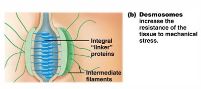
Desmosomes: Anchor cells together, increasing resistance to mechanical stress.
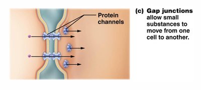
Gap Junctions: Allow small molecules and ions to pass directly between cells for communication.
Classification of Epithelia
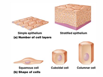
Epithelial tissues are classified by the number of cell layers and the shape of the cells:
Simple Epithelium: One cell layer thick.
Stratified Epithelium: Multiple cell layers.
Cell Shapes: Squamous (flat), cuboidal (cube-shaped), columnar (tall and column-like).
Examples of Epithelial Tissues
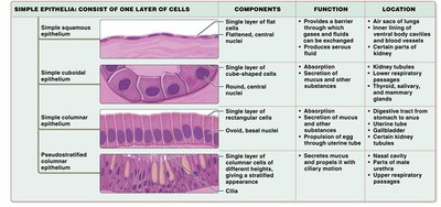
Different types of epithelial tissues are adapted for specific functions and locations in the body.
Simple Squamous Epithelium: Allows for rapid diffusion; found in air sacs of lungs.
Simple Cuboidal Epithelium: Absorption and secretion; found in kidney tubules.
Simple Columnar Epithelium: Absorption and secretion; lines digestive tract.
Pseudostratified Columnar Epithelium: Secretion and movement of mucus; found in respiratory tract.
Stratified and Specialized Epithelia
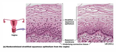
Stratified Squamous Epithelium: Protection; found in skin, mouth, vagina.
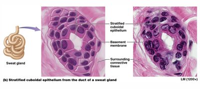
Stratified Cuboidal Epithelium: Protection and secretion; found in sweat gland ducts.
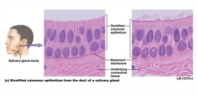
Stratified Columnar Epithelium: Protection and secretion; rare, found in some gland ducts.
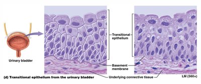
Transitional Epithelium: Stretches and permits distension; lines urinary bladder.
Glandular Epithelia
Glands are structures that secrete products. They are classified as:
Exocrine Glands: Secrete products through ducts to body surfaces or cavities (e.g., sweat, oil glands).
Endocrine Glands: Ductless; secrete hormones directly into the bloodstream, acting at distant sites.
Connective Tissue
Overview and Functions
Connective tissue is the most abundant and widely distributed tissue type. It binds, supports, protects, and transports substances throughout the body.
Binding and Support: Tendons, ligaments, fat, and other structures.
Protection: Fat cushions organs; cartilage protects bones.
Immunity: White blood cells defend against pathogens.
Storage: Fat stores energy; bone stores calcium.
Transport: Blood transports gases, nutrients, and wastes.
Components of Connective Tissue
All connective tissues share three basic components:
Cells: Fibroblasts, adipocytes, immune cells, etc.
Protein Fibers: Collagen (strength), elastic (stretch), reticular (support).
Ground Substance: Gel-like material that fills the space between cells and fibers.
Types of Fibrous Connective Tissue
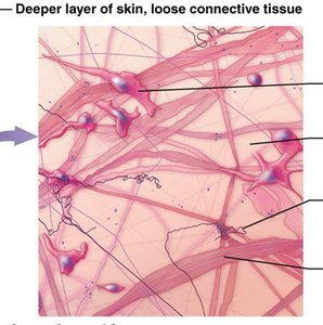
Areolar (Loose) Connective Tissue: Contains fibroblasts, macrophages, mast cells, and white blood cells; supports and binds tissues, holds fluids, and plays a role in inflammation.
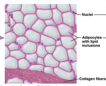
Adipose Tissue: Contains adipocytes (fat cells); stores energy, insulates, and cushions organs.
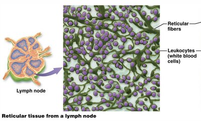
Reticular Tissue: Contains reticular fibers; forms a soft internal skeleton (stroma) supporting other cells in lymphoid organs.
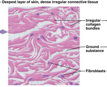
Dense Irregular Connective Tissue: Collagen fibers arranged in multiple directions; provides strength and resists tension in multiple directions; found in dermis and organ capsules.
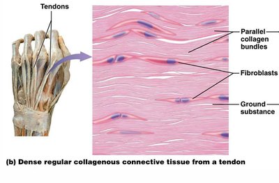
Dense Regular Connective Tissue: Collagen fibers arranged in parallel; provides tensile strength in one direction; found in tendons and ligaments.
Supportive Connective Tissue
Cartilage: Flexible, rubbery matrix; avascular. Types include:
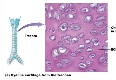
Hyaline Cartilage: Smooth, supports and reinforces; found in joints, ribs, nose, trachea, larynx.
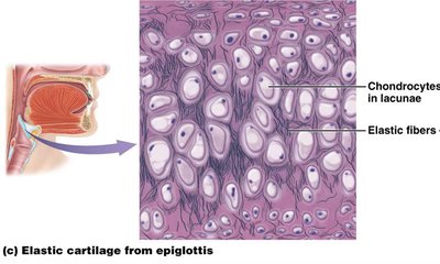
Elastic Cartilage: More elastic fibers; provides flexibility; found in external ear and epiglottis.
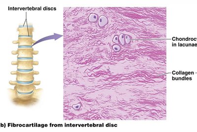
Fibrocartilage: Thick collagen fibers; absorbs shock; found in intervertebral discs, pubic symphysis, knee menisci.
Bone (Osseous Tissue): Rigid matrix; supports and protects, stores calcium, and houses blood-forming cells.
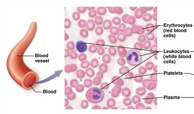
Blood: Fluid connective tissue; transports gases, nutrients, wastes, and other substances.
Muscle Tissue
Types and Functions
Muscle tissue consists of elongated cells specialized for contraction and movement. There are three types:
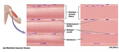
Skeletal Muscle: Voluntary, striated, multinucleate; attached to bones for movement.
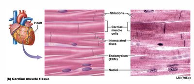
Cardiac Muscle: Involuntary, striated, branched, single nucleus; found only in the heart, connected by intercalated discs.
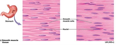
Smooth Muscle: Involuntary, non-striated, spindle-shaped; found in walls of hollow organs (e.g., stomach, blood vessels).
Nervous Tissue
Structure and Function
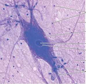
Nervous tissue is specialized for communication by electrical and chemical signals. It consists of two main cell types:
Neurons: Functional units that generate and transmit nerve impulses.
Neuroglial Cells: Support, protect, and nourish neurons.
Summary Table: Major Tissue Types
Tissue Type | Main Function | Key Features | Example Location |
|---|---|---|---|
Epithelial | Protection, secretion, absorption | Cells tightly packed, avascular | Skin, glands, lining of GI tract |
Connective | Support, binding, transport | Cells in matrix of fibers and ground substance | Tendons, fat, blood, bone |
Muscle | Movement | Elongated, contractile cells | Skeletal muscles, heart, walls of hollow organs |
Nervous | Communication | Neurons and supporting cells | Brain, spinal cord, nerves |
