 Back
Human Anatomy & Physiology: Chapter 1 - The Human Body: An Orientation
Study Guide - Smart Notes
 Tailored notes based on your materials, expanded with key definitions, examples, and context.
What is Anatomy and Physiology?
Definitions and Distinctions
Anatomy and physiology are foundational sciences for understanding the human body. Anatomy is the study of the body's structure, while physiology is the study of the body's functions. These disciplines are closely related, as structure often determines function.
Anatomy: Examines the physical structure of organs, tissues, and systems.
Physiology: Investigates how those structures work and interact.
Principle of Complementarity: The function of a body part depends on its structure.
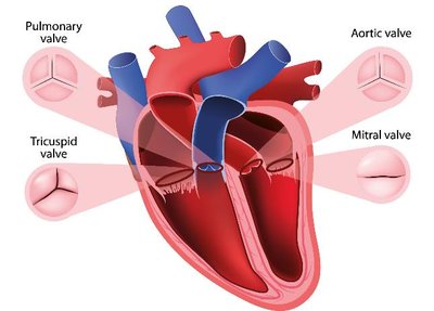
Example: The heart's valves (pulmonary, tricuspid, aortic, mitral) are structured to ensure one-way blood flow, illustrating the relationship between anatomy and physiology.
Levels of Organization
Hierarchy of Structural Complexity
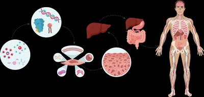
The human body is organized into a hierarchy of levels, each building upon the previous. Changes at one level can affect others.
Atomic and Molecular Level: Atoms and molecules form the basis of all structures.
Macromolecule Level: Large molecules such as proteins and DNA.
Cellular Level: Cells are the basic units of life.
Tissue Level: Groups of similar cells performing a common function.
Organ Level: Structures composed of multiple tissue types.
Organism Level: The complete living individual.
Example: Statins inhibit an enzyme at the molecular level, affecting cholesterol production at the organ level (liver), and ultimately impacting the organism's health.
Variation in Anatomy and Physiology
Normal and Abnormal Variation
There is significant variation in human anatomy and physiology. Reference bodies are used for standardization, but individual differences are common and can impact medical procedures.
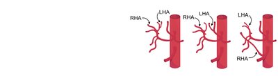
Anatomical Variation: Differences in blood vessel branching, organ position, etc.
Reference Body: Typically a healthy young adult; used for teaching and communication.
Normal Variation: Most variations do not affect function, but extreme variations may cause pathology.
Example: Variations in hepatic artery branching are common, but textbooks present a standard structure for clarity.

Introduction to Organ Systems
Overview of Major Organ Systems
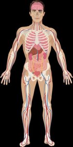
The body is organized into organ systems, each with specific functions. Systems are grouped by their primary roles, but many organs serve multiple functions.
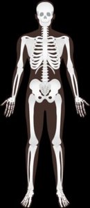
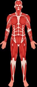
Protection, Structure, & Support: Integumentary, Skeletal, Muscular systems
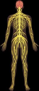
Communication & Integration: Nervous, Endocrine systems
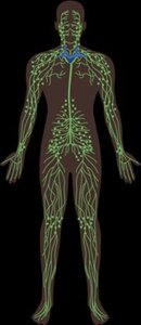
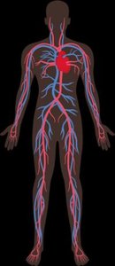
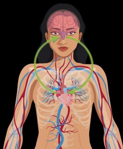
Transport & Immunity: Cardiovascular, Lymphatic systems
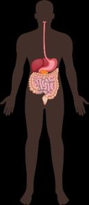
Nutrient, Gas, & Waste Exchange: Respiratory, Digestive, Urinary systems
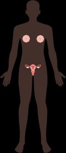
Reproduction: Male and Female Reproductive systems
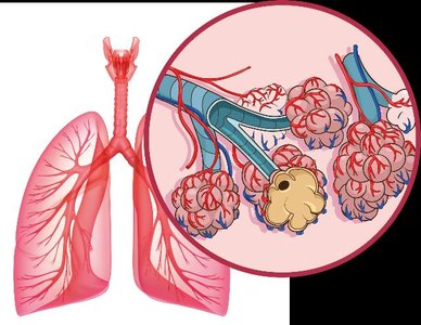
Example: The lungs' structure (alveoli, thin walls, capillaries) enables efficient gas exchange.


Homeostasis
Maintaining Internal Stability
Homeostasis is the maintenance of a stable internal environment. The body regulates variables such as temperature, pH, and blood glucose within narrow ranges.
Set-point: The ideal value for a physiological variable.
Dynamic Equilibrium: Internal conditions fluctuate within a range, not a fixed state.
Failure to maintain homeostasis: Results in pathology (disease).
Example: Blood pH is maintained between 7.35 and 7.45; deviation leads to acidosis or alkalosis.
Feedback Loops
Mechanisms of Homeostatic Control
Homeostasis is regulated by feedback loops:
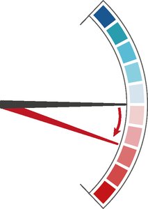
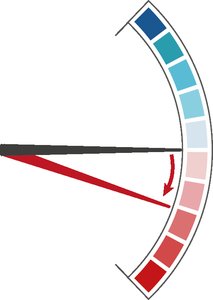
Negative Feedback: Opposes the original stimulus, returning the system to the set-point. Most common.
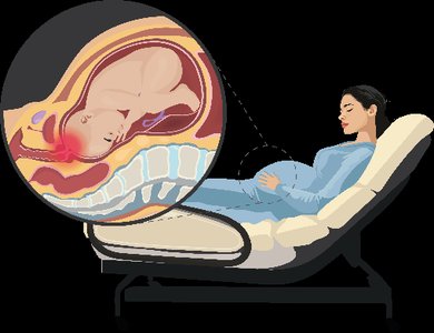
Positive Feedback: Amplifies the original stimulus, moving the system away from the set-point. Less common.
Example: Sweating cools the body (negative feedback); labor contractions increase until birth (positive feedback).












Anatomical Position and Directional Terms
Standard Reference for Describing the Body
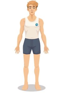
The anatomical position is a universally accepted reference posture for describing locations and directions on the body.
Body upright, face forward, feet together, arms at sides, palms forward.
Left and right refer to the subject's left and right.
Directional Terms: Used to compare locations:
Superior: Toward the head
Inferior: Toward the feet
Anterior (ventral): Toward the front
Posterior (dorsal): Toward the back
Medial: Toward the midline
Lateral: Away from the midline
Proximal: Closer to the point of attachment
Distal: Farther from the point of attachment
Superficial: Closer to the surface
Deep: Further from the surface
Anatomical Terms for Body Regions
Regional Terminology
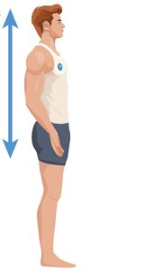
Specific terms are used to describe regions of the body, often derived from Greek or Latin.
Cephalic: Head
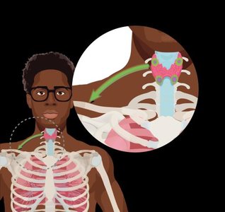
Cervical: Neck
Thoracic: Chest
Abdominal: Abdomen
Pelvic: Pelvis
Inguinal: Groin
Pubic: Genital region
Scapular: Shoulder blade
Lumbar: Lower back
Gluteal: Buttocks
Femoral: Thigh
Patellar: Knee
Crural: Leg
Plantar: Sole of foot
Digital: Fingers/toes
Abdominopelvic Quadrants and Regions
Divisions of the Abdomen and Pelvis
The abdomen and pelvis are divided for clinical and anatomical reference:
Quadrants: Right Upper (RUQ), Left Upper (LUQ), Right Lower (RLQ), Left Lower (LLQ)
Regions: Nine regions (e.g., epigastric, hypogastric, umbilical, lumbar, inguinal, hypochondriac)
Example: The appendix is located in the right lower quadrant; the bladder is in the hypogastric region.
Anatomical Planes and Sections
Planes Used for Dissection and Imaging
Anatomical planes are imaginary lines used to divide the body:
Frontal (coronal): Divides anterior and posterior
Sagittal: Divides left and right (midsagittal = midline, parasagittal = off midline)
Transverse: Divides superior and inferior
Oblique: Divides at an angle
Organization of the Body: Body Cavities
Main Body Cavities
The body contains major cavities that house organs:
Anterior (ventral) cavity: Thoracic and abdominopelvic cavities
Posterior (dorsal) cavity: Cranial and vertebral cavities
Thoracic cavity: Contains heart, lungs, esophagus, trachea
Abdominopelvic cavity: Contains digestive, urinary, and reproductive organs
Serous Membranes
Structure and Function
Serous membranes are double-layered tissues that line body cavities and cover organs. They consist of:
Visceral layer: Attached to the organ
Parietal layer: Attached to the cavity wall
Serous cavity: Space between layers, filled with lubricating serous fluid
Example: The pleura surrounds the lungs; the pericardium surrounds the heart; the peritoneum surrounds abdominal organs.
Summary Table: Major Organ Systems and Functions
System | Main Organs | Primary Function |
|---|---|---|
Integumentary | Skin, hair, nails | Protection, temperature regulation |
Skeletal | Bones, cartilage | Support, protection, movement |
Muscular | Muscles | Movement, heat production |
Nervous | Brain, spinal cord, nerves | Rapid communication, integration |
Endocrine | Glands (pituitary, thyroid, etc.) | Hormonal regulation |
Cardiovascular | Heart, blood vessels | Transport of materials |
Lymphatic | Lymph nodes, vessels | Immunity, fluid balance |
Respiratory | Lungs, trachea | Gas exchange |
Digestive | Stomach, intestines, liver | Nutrient absorption |
Urinary | Kidneys, bladder | Waste removal, fluid balance |
Reproductive | Ovaries, testes | Production of offspring |
Key Equations and Concepts
Blood pH:
Internal Body Temperature:
Blood Glucose: (fasting)
Additional info: These notes provide a comprehensive overview of Chapter 1, suitable for exam preparation and foundational understanding in anatomy and physiology.
