 Back
Human Cardiovascular Physiology: Cardiac Cycle, Heart Sounds, Pulse, and Blood Pressure
Study Guide - Smart Notes
 Tailored notes based on your materials, expanded with key definitions, examples, and context.
Cardiac Cycle
Phases and Events of the Cardiac Cycle
The cardiac cycle describes the sequence of events that occur as the heart beats, including periods of contraction (systole) and relaxation (diastole). Understanding these phases is essential for interpreting heart sounds and blood flow dynamics.
Systole: The phase of contraction, subdivided into atrial systole and ventricular systole.
Diastole: The phase of relaxation, subdivided into atrial diastole and ventricular diastole.
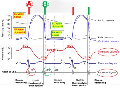
End-Diastolic Volume (EDV): The volume of blood in the ventricles at the end of diastole, just before systole begins.
End-Systolic Volume (ESV): The volume of blood remaining in the ventricles after systole.
Stroke Volume: The amount of blood ejected from the ventricle during systole, calculated as EDV - ESV.
Key Events:
At "A": AV valves close (S1 sound), SL valves open, blood ejection starts.
At "B": SL valves close (S2 sound), AV valves open, ventricular filling starts.
Heart Sounds and Auscultation
Heart Sounds Produced in the Cardiac Cycle
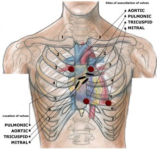
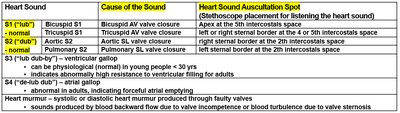
Heart sounds are produced by the closure of heart valves and can be detected using a stethoscope. Auscultation is the process of listening to these sounds at specific anatomical sites.
S1 (lub): Caused by closure of AV valves (bicuspid and tricuspid).
S2 (dub): Caused by closure of semilunar valves (aortic and pulmonary).
S3 and S4: Additional sounds, sometimes heard in specific physiological or pathological conditions.
Stethoscope and Auscultation Technique
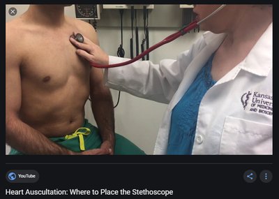
The stethoscope is a precision instrument used for auscultation. Proper placement is crucial for detecting specific valve sounds.
Bell of the stethoscope is placed at designated intercostal spaces to hear S1 and S2 sounds.
Each valve has a specific auscultation site.
Heart Sound Table
Heart Sound | Cause of the Sound | Heart Sound Auscultation Spot |
|---|---|---|
S1 (lub) | Bicuspid AV valve closure | Apex at the 5th intercostal space |
S1 (lub) | Tricuspid AV valve closure | Left sternal border at the 4th intercostal space |
S2 (dub) | Aortic SL valve closure | Right sternal border at the 2nd intercostal space |
S2 (dub) | Pulmonary SL valve closure | Left sternal border at the 2nd intercostal space |
Pulse Palpation
Pulse Points and Palpation Technique
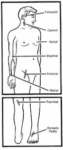
Pulse palpation is the process of feeling the vibration of blood as it passes through arteries. It is performed at peripheral sites where arteries are superficial and cross bones.
Radial artery: At the wrist
Carotid artery: In the neck
Femoral artery: In the groin
Other sites: Temporal, brachial, popliteal, dorsalis pedis
Radial Pulse Measurement
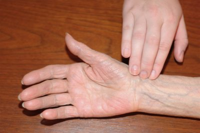
To measure the radial pulse:
Place index and middle fingers on the opposite wrist.
Adjust pressure and position to locate the pulse.
Count beats for 30 seconds and multiply by two for beats per minute.
If rhythm is irregular, count for a full minute.
Palpation vs. Auscultation
Palpation: Feeling the pulse at peripheral arteries.
Auscultation: Listening to heart sounds at central sites near the heart.
Blood Pressure
Arterial Blood Pressure Concepts
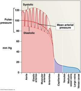
Blood pressure is the force exerted by circulating blood on the walls of arteries. It is measured in millimeters of mercury (mmHg) and consists of several key values:
Systolic pressure: Peak pressure during ventricular systole.
Diastolic pressure: Minimum pressure during diastole.
Pulse pressure: Difference between systolic and diastolic pressure.
Mean arterial pressure (MAP): Average pressure in the arteries, calculated as:
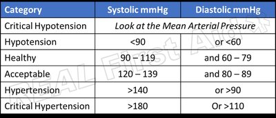
Blood Pressure Categories
Blood pressure is classified into categories based on systolic and diastolic values.
Category | Systolic mmHg | Diastolic mmHg |
|---|---|---|
Critical Hypotension | Look at the Mean Arterial Pressure | |
Hypotension | <90 | or <60 |
Healthy | 90–119 | and 60–79 |
Acceptable | 120–139 | and 80–89 |
Hypertension | >140 | or >90 |
Critical Hypertension | >180 | or >110 |
Blood Pressure Measurement: Principle and Procedure
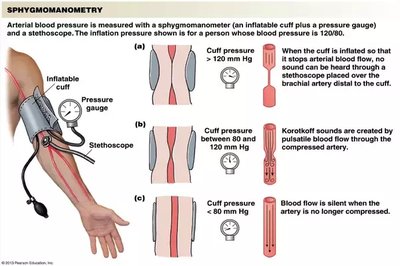
Blood pressure is measured using a sphygmomanometer and a stethoscope, typically at the brachial artery. The procedure involves inflating a cuff and listening for Korotkoff sounds.
Inflate cuff above systolic pressure: Brachial artery is occluded, no sound is heard.
Release cuff to systolic pressure: Blood spurts through the constricted artery, producing Korotkoff sounds (onset).
Release cuff to diastolic pressure: Artery opens fully, sounds disappear (offset).
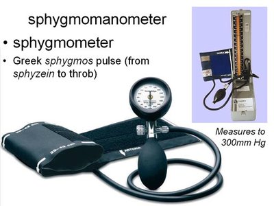
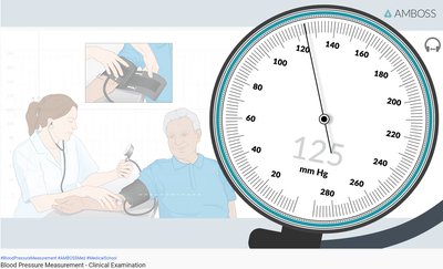
Devices Used in Blood Pressure Measurement
Sphygmomanometer: Inflatable cuff and pressure gauge.
Stethoscope: Used to detect Korotkoff sounds.
Blood Flow and Korotkoff Sounds
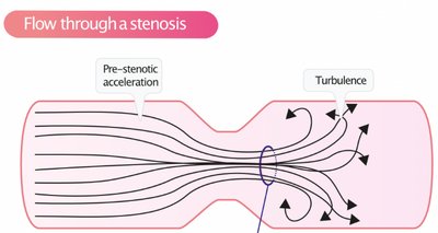
Korotkoff sounds are produced by turbulent blood flow through a constricted artery.
Above systolic pressure: No sound (artery closed).
At systolic pressure: Onset of Korotkoff sounds (turbulent flow).
At diastolic pressure: Offset of Korotkoff sounds (artery open, laminar flow).
Review Questions and Answers
The “1” position is the best spot to hear Aortic S2 sound.
If a person has a blood pressure of 120/90, his mean arterial pressure would be 100 mm Hg.
Onset of Korotkoff sounds represents Systolic pressure.
The cause of S1 heart sound is The AV valves closure.
Summary Table: Key Terms and Definitions
Term | Definition |
|---|---|
Cardiac Cycle | Sequence of events in one heartbeat, including systole and diastole |
EDV | End-diastolic volume; blood in ventricle at end of diastole |
ESV | End-systolic volume; blood in ventricle at end of systole |
Stroke Volume | EDV minus ESV; blood ejected per beat |
Systolic Pressure | Peak arterial pressure during ventricular contraction |
Diastolic Pressure | Minimum arterial pressure during relaxation |
Pulse Pressure | Difference between systolic and diastolic pressure |
Mean Arterial Pressure | Average arterial pressure; MAP = Diastolic + 1/3 Pulse Pressure |
Korotkoff Sounds | Sounds heard during blood pressure measurement, caused by turbulent flow |
Auscultation | Listening to heart sounds with a stethoscope |
Palpation | Feeling the pulse at peripheral arteries |
