 Back
BackHuman Development: From Fertilization to Birth
Study Guide - Smart Notes
 Tailored notes based on your materials, expanded with key definitions, examples, and context.
module 10 part 1
Human Development
Introduction to Human Development
Human development is the process by which a single fertilized cell transforms into a complex multicellular organism. This process encompasses the stages from fertilization to birth, including pre-embryonic, embryonic, and fetal development. The gestation period, typically 280 days from the last menstrual period, is divided into these stages based on the developmental milestones of the conceptus.
Pregnancy: The period from fertilization to birth.
Conceptus: The developing offspring during the pre-embryonic, embryonic, and fetal stages.
Gestation period: The time from the last menstrual period to birth (about 280 days).
Embryo: The conceptus from the third to the eighth week after fertilization.
Fetus: The conceptus from the ninth week until birth.
From Egg to Embryo: Accomplishing Fertilization
Sperm Transport and Capacitation
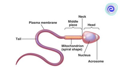
Fertilization requires the sperm to reach and penetrate the ovulated secondary oocyte. The journey is challenging due to the acidic vaginal environment, cervical mucus, and immune defenses in the uterus. Only a small fraction of sperm reach the uterine tubes, and they must undergo capacitation—a process that makes their membranes fragile enough to release acrosomal enzymes necessary for oocyte penetration.
Capacitation: The process by which sperm membranes become fragile, allowing the release of hydrolytic enzymes from the acrosome.
Acrosome: A cap-like structure on the sperm head containing enzymes for oocyte penetration.
Acrosomal Reaction and Sperm Penetration
The acrosomal reaction is the release of enzymes that digest the protective layers around the oocyte (corona radiata and zona pellucida). Hundreds of sperm contribute to this process, but only one will fertilize the oocyte by fusing its membrane and delivering its nucleus into the oocyte cytoplasm.
Blocks to Polyspermy
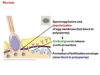
To ensure monospermy (entry of only one sperm), two mechanisms prevent polyspermy:
Fast block: Depolarization of the oocyte membrane upon sperm fusion, preventing further sperm entry.
Slow block (Cortical reaction): Release of cortical granules causes changes in the zona pellucida, detaching other sperm and establishing a permanent block.
Completion of Meiosis II and Fertilization
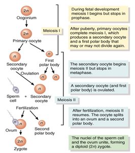
After sperm entry, the secondary oocyte completes meiosis II, forming the ovum and a second polar body. The fusion of maternal and paternal chromosomes produces the diploid zygote, marking the true act of fertilization.
Pre-embryonic Development
Cleavage and Blastocyst Formation
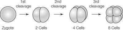
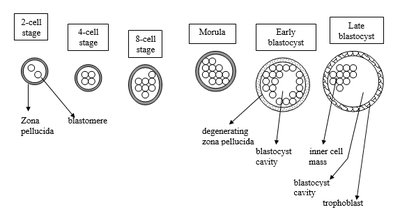
Cleavage is a series of rapid mitotic divisions of the zygote, resulting in smaller cells called blastomeres. This process increases the surface-to-volume ratio, facilitating nutrient uptake and waste removal. The conceptus progresses from a zygote to a morula and then to a blastocyst.
Blastomeres: The smaller cells produced during cleavage.
Morula: A solid ball of cells formed after several cleavage divisions.
Blastocyst: A hollow sphere with an outer trophoblast layer and an inner cell mass.
Implantation
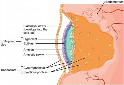
Implantation begins about six days after ovulation when the blastocyst adheres to the endometrium. The trophoblast differentiates into two layers: the cytotrophoblast (retains cell boundaries) and the syncytiotrophoblast (multinucleated mass). The trophoblast secretes enzymes to facilitate implantation and produces human chorionic gonadotropin (HCG) to maintain the corpus luteum and prevent menstruation.
Placentation
Formation and Structure of the Placenta
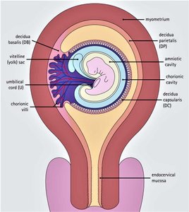
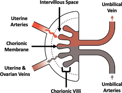
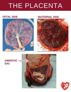
Placentation is the formation of the placenta, a temporary organ derived from both embryonic and maternal tissues. The chorion develops fingerlike chorionic villi, which become vascularized and connect to the embryo via the umbilical arteries and vein. The placenta functions as a nutritive, respiratory, excretory, and endocrine organ.
Chorion: The outermost fetal membrane, forming the fetal part of the placenta.
Chorionic villi: Projections that increase surface area for exchange between maternal and fetal blood.
Decidua basalis: The maternal part of the placenta.
Decidua capsularis: Endometrial cells surrounding the embryo on the uterine cavity side.
Placental Hormones and Clinical Considerations
The placenta secretes HCG early in pregnancy, followed by estrogen and progesterone. These hormones maintain the endometrium and support mammary gland development. Teratogens (e.g., alcohol, nicotine, certain drugs, infections) can cross the placenta and cause congenital abnormalities.
Fetal Alcohol Syndrome (FAS): Caused by maternal alcohol consumption, leading to microcephaly and developmental delays.
Nicotine: Reduces oxygen delivery, resulting in low birth weight.
Embryonic Development
Gastrulation and Germ Layer Formation
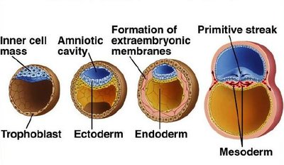
Gastrulation transforms the blastocyst's inner cell mass into a three-layered embryo (gastrula) with ectoderm, mesoderm, and endoderm. The primitive streak forms, establishing the embryo's longitudinal axis. The notochord, derived from mesoderm, provides initial axial support.
Ectoderm: Forms the nervous system and skin epidermis.
Endoderm: Forms the mucosal linings of the digestive and respiratory tracts.
Mesoderm: Forms muscles, bones, and most other tissues.
Embryonic Membranes
Four embryonic membranes form during early development: amnion, yolk sac, chorion, and allantois. The amnion provides a protective, fluid-filled environment. Amniotic fluid is initially derived from maternal blood and later includes fetal urine. Amniocentesis can sample this fluid for genetic testing.
Organogenesis
Organogenesis is the process by which the three germ layers differentiate into the body's organs and systems. By the end of the embryonic period (week 8), all major organ systems are established, and the embryo is about 22 mm long.
Ossification of bones begins.
Skeletal muscles start contracting.
Kidneys, gonads, lungs, and digestive organs reach advanced development.
Heart and liver are prominent and functional.
Sex Determination
The testis-determining factor (TDF) gene initiates male sex differentiation. In its absence, the embryo develops as female. TDF acts as a transcription factor, promoting the formation of seminiferous tubules and subsequent testosterone production.
Prenatal Circulation and Birth Adaptations
Prenatal Circulatory Pattern
The fetal circulatory system includes unique structures that bypass nonfunctional organs (lungs, liver):
Umbilical vein: Carries oxygenated blood from the placenta to the fetus.
Ductus venosus: Shunts blood past the liver to the inferior vena cava.
Foramen ovale: Allows blood to flow from the right to the left atrium, bypassing the lungs.
Ductus arteriosus: Connects the pulmonary trunk to the aorta, further bypassing the lungs.
Umbilical arteries: Return deoxygenated blood to the placenta.
Cardiovascular Changes at Birth
At birth, the newborn must transition to independent life. The first breath inflates the lungs, and circulatory shunts close:
Umbilical vessels constrict and become ligaments.
Ductus venosus becomes the ligamentum venosum.
Foramen ovale closes, forming the fossa ovalis.
Ductus arteriosus becomes the ligamentum arteriosum.
These changes ensure that blood now circulates through the lungs and liver, supporting the newborn's independent respiration and metabolism.
