 Back
BackHuman Development: From Fertilization to Birth
Study Guide - Smart Notes
 Tailored notes based on your materials, expanded with key definitions, examples, and context.
Human Development
Introduction to Human Development
Human development is the process of progressive changes that occur from the fertilization of an egg to maturity and ultimately to death. This process involves a series of well-coordinated events that transform a single cell into a complex multicellular organism.
Development includes cellular division, differentiation, and morphogenesis.
Genetic information is halved during gamete formation and restored at fertilization.
Fertilization and Early Development
Penetration and Fertilization
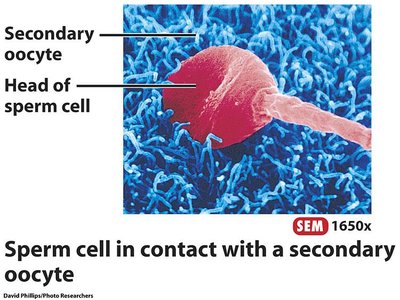
Fertilization is the union of a sperm cell and a secondary oocyte, resulting in the formation of a zygote. The sperm must penetrate several layers to reach the oocyte's cytoplasm.
Corona radiata: Outer layer of follicular cells surrounding the oocyte.
Zona pellucida: Glycoprotein layer beneath the corona radiata.
Plasma membrane of secondary oocyte: The sperm must fuse with this membrane to deliver its genetic material.
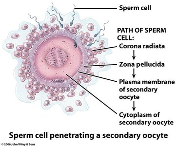
Upon entry of the sperm, the secondary oocyte completes meiosis II, casting out the second polar body. Fertilization is complete when the male and female pronuclei unite, restoring the diploid chromosome number.
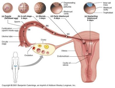
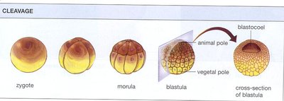
Preembryonic Development: Cleavage, Morula, and Blastocyst Formation
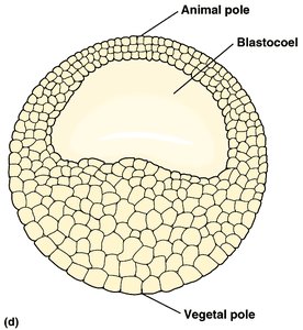
After fertilization, the zygote undergoes rapid mitotic divisions called cleavage, forming a solid ball of cells known as the morula. The morula then develops into a hollow structure called the blastocyst (or blastula in non-mammalian vertebrates), which contains a fluid-filled cavity called the blastocoel.
Zygote: Single fertilized cell.
Morula: Solid ball of cells resulting from cleavage.
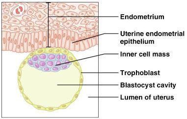
Blastocyst: Hollow sphere with an inner cell mass and outer trophoblast layer.

Phases of Embryonic Development
Cleavage
Cleavage is the series of rapid mitotic divisions that increase the number of cells without increasing the overall size of the embryo. This process produces the morula and then the blastula.
Cleavage partitions the cytoplasm of the large zygote into many smaller cells.
The blastocoel forms within the blastula, providing a cavity for further development.
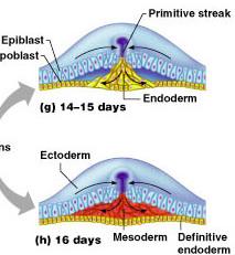
Gastrulation
Gastrulation is the process by which the blastula reorganizes into a three-layered structure called the gastrula. Each layer, known as a germ layer, will give rise to specific tissues and organs.
Ectoderm: Forms the epidermis and nervous system.
Mesoderm: Forms muscles, skeleton, and circulatory system.
Endoderm: Forms the digestive tract and associated organs.
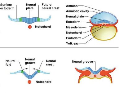
Organogenesis
Organogenesis is the phase during which the three germ layers develop into the internal organs of the organism. The first major event is the formation of the notochord and the neural tube (neurulation), which will become the central nervous system.
Notochord: Temporary rod-shaped structure that provides support and induces neural development.
Neural plate and tube: Ectodermal cells thicken and fold to form the neural tube, precursor to the brain and spinal cord.
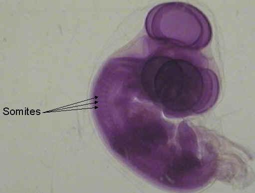
Somites: Segmented blocks of mesoderm that give rise to vertebrae and skeletal muscles.
Human Embryonic and Fetal Development
Timeline of Early Human Development
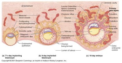
Week 1: Fertilization, cleavage, morula, blastocyst formation, and implantation in the uterus.
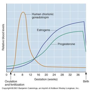
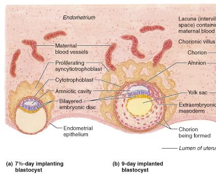
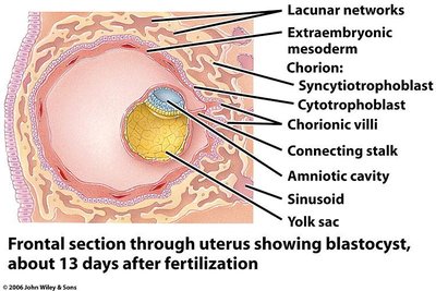
Week 2: Gastrulation and formation of germ layers; trophoblast secretes hCG to maintain the corpus luteum.
Week 3-4: Beginning of organ development, neural tube formation, heart development, and limb bud appearance.
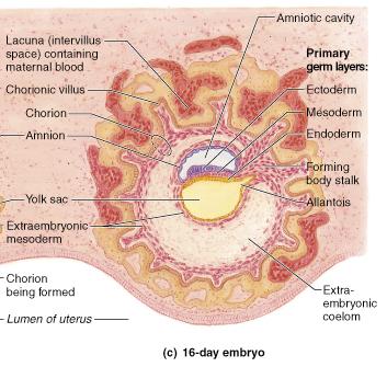
Placentation and Embryonic Membranes
The placenta forms from the chorion and maternal tissues, facilitating nutrient and gas exchange between mother and fetus. The embryo is supported by three main external membranes: chorion, amnion, and allantois.
Chorion: Develops fingerlike villi that increase surface area for exchange.
Amnion: Encloses the embryo in a fluid-filled cavity for protection.
Allantois: Forms part of the umbilical cord and contributes to blood vessel development.
Embryonic and Fetal Growth
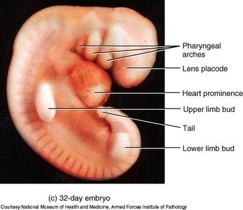
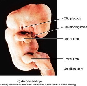
By the end of the embryonic period (8 weeks), all major organ systems are established. The fetal period (weeks 9 to birth) is characterized by growth and refinement of these systems.
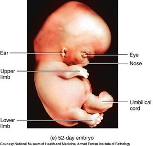
Week 6-8: Embryo is recognizable as human; organ systems are established.
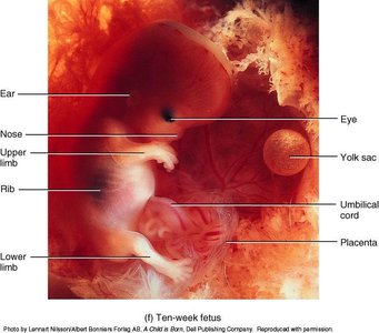
10 weeks: Fetus continues to grow; placenta fully functional.
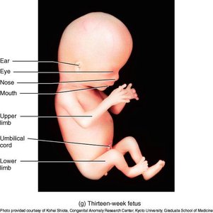
13 weeks: Further development of limbs and facial features.
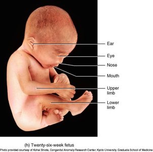
26 weeks: Continued growth and maturation of organ systems.
Maternal Changes During Pregnancy
Physiological Adaptations
Pregnancy induces significant changes in the mother's body to support fetal development.
Relaxin: Hormone that relaxes pelvic ligaments and the pubic symphysis.
Weight gain: Average of 29 pounds.
Gastrointestinal: Morning sickness due to elevated estrogen and progesterone.
Urinary: Increased urine production to handle fetal wastes.
Respiratory: Nasal congestion and possible dyspnea.
Cardiovascular: Increased blood volume (25-40%), possible varicose veins due to impaired venous return.
Lactation
Advantages of Breast Milk
Breastfeeding provides numerous benefits for both the infant and the mother.
Nutritional: Fats and iron are better absorbed; amino acids are efficiently metabolized.
Immunological: Contains IgA, other immunoglobulins, complement, lysozyme, interferon, and lactoperoxidase.
Developmental: Promotes neurological, jaw, teeth, and facial development; supports speech development.
Maternal health: Reduces risks for breast and ovarian cancer; strengthens maternal-child bond.
Breast milk also contains interleukins and prostaglandins to prevent excessive inflammatory responses and natural laxatives to help cleanse the infant's bowels of meconium.
