Back
BackHuman Development: Prenatal Period, Pregnancy, and Postnatal Changes
Study Guide - Smart Notes
 Tailored notes based on your materials, expanded with key definitions, examples, and context.
Development and the Prenatal Period
Overview of the Prenatal Period
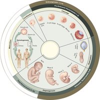
The prenatal period encompasses the time from fertilization to birth, lasting approximately 38 weeks. It is divided into three main stages: the pre-embryonic period (first 2 weeks), the embryonic period (weeks 3–8), and the fetal period (weeks 9–38). Each stage is characterized by specific developmental milestones and changes in the structure and function of the developing organism.
Pre-embryonic period: Fertilization to implantation; formation of the blastocyst.
Embryonic period: Major organ systems begin to develop; the organism is termed an embryo.
Fetal period: Growth and maturation of tissues and organs; the organism is termed a fetus.
Fertilization and Early Development
Fertilization
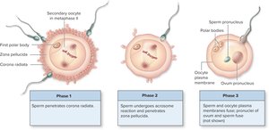
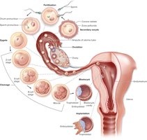
Fertilization is the process by which two gametes (sperm and secondary oocyte) fuse to form a diploid zygote. This event restores the diploid chromosome number and determines the genetic sex of the offspring. Fertilization typically occurs in the ampulla of the uterine tube.
Capacitation: Sperm undergo physiological changes in the female reproductive tract to become capable of fertilizing the oocyte.
Phases of fertilization:
Penetration of the corona radiata
Penetration of the zona pellucida (acrosome reaction)
Fusion of sperm and oocyte plasma membranes and pronuclei
Prevention of polyspermy: Hardening of the zona pellucida ensures only one sperm fertilizes the oocyte.
Cleavage and Blastocyst Formation
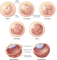
After fertilization, the zygote undergoes a series of rapid mitotic divisions called cleavage. These divisions increase the cell number without increasing the overall size of the structure. The process leads to the formation of a morula (16-cell stage) and then a blastocyst, which consists of an outer trophoblast and an inner embryoblast.
Morula: Solid ball of cells resulting from cleavage.
Blastocyst: Fluid-filled structure with a trophoblast (forms chorion) and embryoblast (forms embryo proper).
Pluripotency: Embryoblast cells can differentiate into any tissue type.
Transit Through the Uterine Tube and Implantation
The pre-embryo travels through the uterine tube toward the uterus, undergoing cleavage and forming the blastocyst. By the end of the first week, the blastocyst enters the uterine lumen, sheds the zona pellucida, and begins implantation into the endometrial lining.
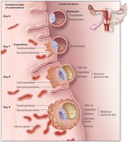
Implantation: The blastocyst burrows into the endometrium, beginning around day 7 post-fertilization.
Trophoblast differentiation: Forms the cytotrophoblast (inner layer) and syncytiotrophoblast (outer layer).
Human chorionic gonadotropin (hCG): Produced by syncytiotrophoblast; maintains corpus luteum and is the basis for pregnancy tests.
Hormonal Changes During Early Pregnancy
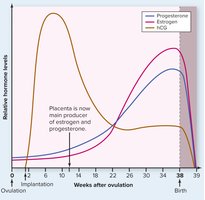
Hormone levels fluctuate significantly during pregnancy. hCG is produced early to maintain the corpus luteum, while estrogen and progesterone levels rise to support the uterine lining and fetal development. The placenta eventually takes over hormone production.
hCG: Peaks early, then declines as the placenta assumes hormone production.
Estrogen and progesterone: Essential for maintaining pregnancy and preparing the body for childbirth.
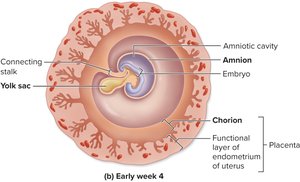
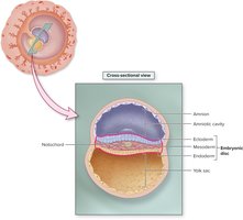
Formation of Extraembryonic Membranes and Placenta
Bilaminar Germinal Disc and Extraembryonic Membranes
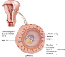
By day 8, the embryoblast forms two layers: the hypoblast and epiblast, together called the bilaminar germinal disc. Extraembryonic membranes develop to protect and support the embryo.
Yolk sac: Early site of blood cell formation.
Amnion: Encloses the embryo in a fluid-filled sac for protection.
Chorion: Outermost membrane; forms the fetal portion of the placenta.
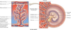
Placenta Development and Function
The placenta is a highly vascular organ that facilitates the exchange of nutrients, gases, and wastes between maternal and fetal blood. It also produces hormones necessary for pregnancy maintenance.
Chorionic villi: Fingerlike projections containing fetal blood vessels for exchange with maternal blood.
Selective permeability: Allows passage of gases and nutrients but blocks many harmful substances.
Placental hormones: Estrogen, progesterone, and hCG.
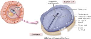
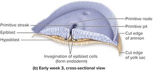
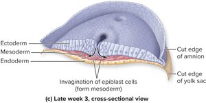
Gastrulation and Formation of Germ Layers
Gastrulation
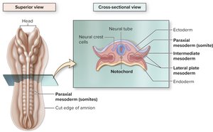
Gastrulation is a critical process in the third week of development, forming the three primary germ layers from the epiblast: ectoderm, mesoderm, and endoderm. These layers give rise to all tissues and organs of the body.
Primitive streak: Site of cell migration and germ layer formation.
Ectoderm: Forms nervous system and skin.
Mesoderm: Forms muscle, bone, and connective tissue.
Endoderm: Forms internal linings of organs.
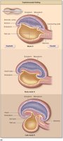
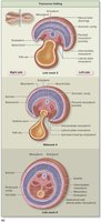
Folding of the Embryonic Disc and Differentiation
Embryonic Folding
During the late third and fourth weeks, the embryonic disc undergoes cephalocaudal and transverse folding, transforming the flat disc into a cylindrical embryo. This process establishes the basic body plan.
Cephalocaudal folding: Head and tail regions fold toward each other.
Transverse folding: Lateral edges move ventrally, enclosing the gut tube.
Differentiation of Germ Layers
Each germ layer differentiates into specific tissues and organs:
Ectoderm: Epidermis, nervous system, sense organs, enamel of teeth.
Mesoderm: Muscle, bone, cardiovascular system, kidneys, reproductive organs.
Endoderm: Lining of gastrointestinal, respiratory, and urinary tracts; liver, pancreas.
Organogenesis and the Fetal Period
Organogenesis
Organogenesis is the process of organ formation, beginning after germ layer differentiation and embryonic folding. By the end of the eighth week, most organ systems have a rudimentary form. This period is highly sensitive to teratogens, which can cause birth defects.
Fetal Period
The fetal period extends from the beginning of the third month to birth. It is characterized by rapid growth, tissue maturation, and further development of organ systems. The fetus increases dramatically in size and weight, especially in the last two months of gestation.
Pregnancy: Maternal Changes and Hormonal Regulation
Hormonal Changes
Pregnancy involves significant hormonal changes to support fetal development and prepare the mother's body for childbirth and lactation.
Estrogen and progesterone: Maintain uterine lining, suppress ovarian cycle, promote breast development.
hCG: Maintains corpus luteum early in pregnancy.
Relaxin: Increases flexibility of pelvic ligaments.
Human placental lactogen (HPL): Alters maternal metabolism to provide nutrients for the fetus.
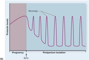
Prolactin: Prepares mammary glands for milk production.
Oxytocin: Stimulates uterine contractions and milk ejection.
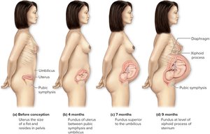
Uterine and Breast Changes
The uterus enlarges significantly during pregnancy, displacing abdominal organs and affecting urinary and digestive systems. Breasts become tender and increase in size due to hormonal stimulation and preparation for lactation.
Labor and Childbirth
Initiation and Stages of Labor
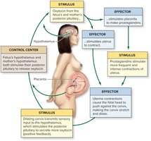
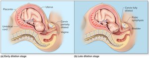
Labor is the process of expelling the fetus and placenta from the uterus, typically at 38 weeks for a full-term pregnancy. True labor is characterized by regular, intense uterine contractions and cervical changes, initiated by a positive feedback loop involving oxytocin and prostaglandins.
Dilation stage: Cervix dilates to 10 cm; amniotic sac ruptures.
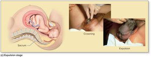
Expulsion stage: Fetus is delivered through the birth canal.
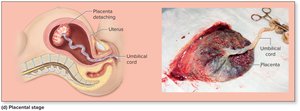
Placental stage: Placenta and fetal membranes are expelled (afterbirth).
Postnatal Changes and Lactation
Hormonal and Fluid Changes
After delivery, estrogen and progesterone levels drop rapidly, leading to postpartum physiological changes. Excess fluid is eliminated through urination, sweating, and lochia (vaginal discharge).
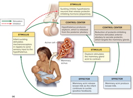
Lactation
Lactation is the production and release of breast milk, regulated by prolactin (milk production) and oxytocin (milk ejection). The process is maintained by a positive feedback mechanism initiated by infant suckling.
Colostrum: First milk produced, rich in immunoglobulins.
Breast milk: Optimal nutrition for the infant, containing fats, proteins, and antibodies.
Inhibition of ovulation: Regular breastfeeding suppresses GnRH, reducing FSH and LH secretion.
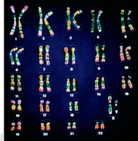
Human Genetics Overview
Chromosomes and Heredity
Humans have 23 pairs of chromosomes, including 22 pairs of autosomes and one pair of sex chromosomes (XX or XY). Genes located on these chromosomes determine inherited traits. Chromosomes are visualized as a karyotype, which can be used to detect chromosomal abnormalities.