 Back
Human Special Senses: Olfaction, Gustation, Vision, Hearing, and Balance
Study Guide - Smart Notes
 Tailored notes based on your materials, expanded with key definitions, examples, and context.
Special Senses Overview
Introduction to Special Senses
The human body possesses specialized senses that allow us to perceive the environment through smell, taste, vision, hearing, and balance. These senses rely on unique receptor mechanisms and neural pathways to transmit information to the brain for interpretation. Perception is influenced by the state of the nervous system, including sympathetic activation and the ability to concentrate or tune out certain sensory signals.
Perception requires both receptor activation and sensory information processing.
Receptors are located in sense organs such as the eye, ear, nose, and tongue.
Sensory information is distributed to specific areas of the cerebral cortex for interpretation.
Olfaction: Sense of Smell
Anatomy and Physiology of Olfaction
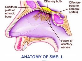
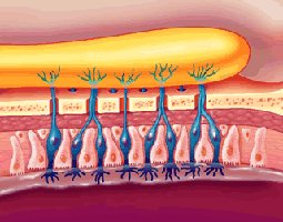
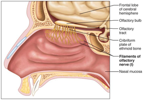
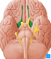
Olfaction is the sense of smell, mediated by specialized olfactory tissue in the nasal cavity. Olfactory receptors are dendrites of neurons that generate action potentials when stimulated by airborne molecules. The olfactory bulbs, located above the cribiform plate, transmit signals to the cerebral cortex via the olfactory tract.
Olfactory epithelium contains receptor cells, supporting cells, and basal cells.
Olfactory bulbs are masses of nerve cells above the cribiform plate of the ethmoid bone.
Olfactory tract carries information to the cerebral cortex for processing.
Receptors are activated by odorant molecules, leading to nerve impulses sent to the brain.
Olfactory Pathways
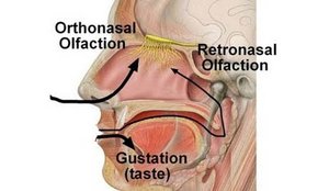
Olfactory information is processed through two main pathways: orthonasal (through the nostrils) and retronasal (from the oral cavity during eating). Both pathways contribute to the perception of flavor and odor.
Orthonsal olfaction: Odorants enter through the nostrils.
Retronasal olfaction: Odorants enter from the oral cavity during eating.
Gustation: Sense of Taste
Anatomy and Physiology of Gustation
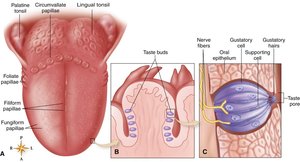
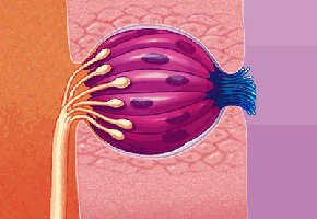
Gustation is the sense of taste, mediated by taste buds located on the tongue and other areas of the oral cavity. Taste buds contain receptor cells that respond to dissolved food molecules, generating nerve impulses sent to the brain.
Taste buds are clusters of receptor cells found in papillae on the tongue.
Each taste bud contains 40-100 receptor cells.
Primary taste sensations include sweet, salty, sour, and bitter.
Food molecules must dissolve in saliva to stimulate taste receptors.
Taste Sensations and Pathways
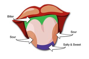
The tongue is divided into regions that are more sensitive to specific taste sensations. Taste signals are transmitted via cranial nerves to the gustatory cortex for interpretation.
Sweet and salty: Detected primarily at the tip and sides of the tongue.
Sour: Detected at the sides of the tongue.
Bitter: Detected at the back of the tongue.
Vision: Anatomy and Physiology of the Eye
Structure of the Human Eye
The human eye is a spherical organ recessed in the bony orbit of the skull. It consists of three main layers: fibrous tunic, vascular tunic, and neural tunic (retina). Accessory structures include eyelids, eyebrows, eyelashes, lacrimal glands, and extrinsic eye muscles.
Fibrous tunic: Outermost layer, includes the cornea (transparent) and sclera (white, opaque).
Vascular tunic: Middle layer, includes the iris (controls pupil size), ciliary body (adjusts lens shape), and choroid (supplies nutrients).
Neural tunic (retina): Innermost layer, contains photoreceptors (rods and cones).
Optics and Visual Processing
Light enters the eye through the cornea and lens, forming an image on the retina. Photoreceptors transform light into nerve signals, which are sent to the visual centers of the brain via the optic nerve.
Rods: Sensitive to light and dark, shapes, and movement; concentrated at the periphery of the retina.
Cones: Sensitive to color (red, green, blue); concentrated in the fovea centralis for sharp vision.
Optic disc: Area where the optic nerve enters the eyeball; lacks photoreceptors (blind spot).
Hearing: Anatomy and Physiology of the Ear
Structure of the Human Ear
The ear is an organ of both hearing and balance, consisting of three main parts: external (outer) ear, middle ear, and internal (inner) ear. Each part plays a distinct role in sound perception and equilibrium.
External ear: Includes the auricle, external auditory canal, and tympanic membrane (eardrum).
Middle ear: Air-filled cavity housing auditory ossicles (malleus, incus, stapes) and the auditory tube.
Inner ear: Contains the cochlea (hearing) and vestibular organs (balance).
Sound Perception
Sound waves are collected by the auricle and directed into the external auditory canal, causing the tympanic membrane to vibrate. Vibrations are transmitted through the auditory ossicles to the cochlea, where hair cells generate action potentials sent to the auditory cortex.
Tympanic membrane: Converts sound waves to mechanical vibrations.
Auditory ossicles: Transmit vibrations to the inner ear.
Cochlea: Contains hair cells that transform vibrations into nerve signals.
Balance: Vestibular System
Anatomy and Physiology of Balance
The vestibular system in the inner ear is responsible for the perception of body position and movement. It includes the semicircular canals, utricle, and saccule, which detect rotational and linear movements.
Semicircular canals: Detect rotational movements.
Utricle: Detects horizontal movements.
Saccule: Detects vertical movements.
Fluid movement bends stereocilia, generating action potentials sent to the brain via the vestibular nerve.
The brain interprets somatosensory information, allowing awareness of body position and movement.
Summary Table: Special Senses and Their Functions
Sense | Receptor Organ | Primary Function |
|---|---|---|
Olfaction | Nasal cavity (olfactory epithelium) | Detection of airborne chemicals (smell) |
Gustation | Tongue (taste buds) | Detection of dissolved chemicals (taste) |
Vision | Eye (retina) | Detection of light and color (sight) |
Hearing | Ear (cochlea) | Detection of sound waves (hearing) |
Balance | Ear (vestibular system) | Detection of body position and movement |
Additional info: Academic context was added to clarify the structure and function of each special sense, including definitions, examples, and pathways. The notes are structured to provide a comprehensive overview suitable for ANP college students preparing for exams.
