Back
BackHuman Structural Architecture & Biomechanics: Bones, Joints, and Movement
Study Guide - Smart Notes
 Tailored notes based on your materials, expanded with key definitions, examples, and context.
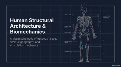
Human Structural Architecture & Biomechanics
Overview
This section introduces the foundational concepts of human skeletal structure, focusing on osseous tissue, skeletal geography, and the mechanics of articulation. Understanding these principles is essential for grasping how the body supports movement, protects organs, and maintains structural integrity.
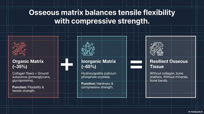
Osseous Tissue Composition
Organic and Inorganic Matrix
Bone tissue is a composite material, balancing flexibility and strength through its organic and inorganic components:
Organic Matrix (~35%): Composed of collagen fibers and ground substance (proteoglycans, glycoproteins). Function: Provides flexibility and tensile strength.
Inorganic Matrix (~65%): Primarily hydroxyapatite (calcium phosphate crystals). Function: Provides hardness and compressive strength.
Resilient Osseous Tissue: The combination of both matrices results in bone that is both strong and flexible. Without collagen, bone is brittle; without minerals, bone bends.
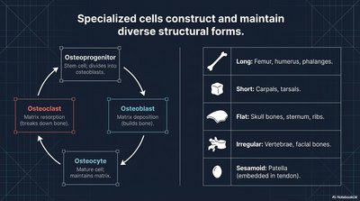
Bone Cells and Structural Forms
Cell Types and Bone Classification
Specialized cells construct and maintain bone tissue, while bones are classified by shape and function:
Osteoprogenitor: Stem cells that differentiate into osteoblasts.
Osteoblast: Responsible for bone matrix deposition (building bone).
Osteocyte: Mature bone cells that maintain the matrix.
Osteoclast: Responsible for bone resorption (breaking down bone).
Long Bones: Femur, humerus, phalanges.
Short Bones: Carpals, tarsals.
Flat Bones: Skull bones, sternum, ribs.
Irregular Bones: Vertebrae, facial bones.
Sesamoid Bones: Patella (embedded in tendon).
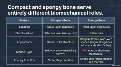
Compact vs. Spongy Bone
Biomechanical Roles and Structure
Compact and spongy bone serve distinct structural and functional roles:
Feature | Compact Bone | Spongy Bone |
|---|---|---|
Location | Outer layer, diaphysis | Inner layer, epiphyses |
Structural Unit | Osteon (Haversian system) | Trabeculae |
Appearance | Dense, solid structure | Irregular lattice-work with spaces |
Marrow Type | Yellow marrow (medullary cavity) | Red marrow (between trabeculae) |
Primary Function | Strength, protection | Shock absorption, houses red marrow |
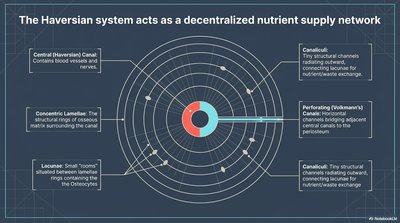
The Haversian System (Osteon)
Decentralized Nutrient Supply
The osteon is the fundamental unit of compact bone, facilitating nutrient and waste exchange:
Central (Haversian) Canal: Contains blood vessels and nerves.
Concentric Lamellae: Rings of osseous matrix surrounding the canal.
Lacunae: Small spaces housing osteocytes.
Canaliculi: Tiny channels connecting lacunae for nutrient/waste exchange.
Perforating (Volkmann's) Canals: Connect adjacent central canals.
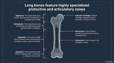
Long Bone Structure
Specialized Zones
Long bones are organized into regions with distinct functions:
Epiphysis: Expanded ends, spongy bone covered by compact bone.
Metaphysis: Transitional zone containing the epiphyseal plate (growth plate in children).
Diaphysis: Central shaft, thick compact bone enclosing the medullary cavity (yellow marrow).
Articular Cartilage: Hyaline cartilage capping joint surfaces to reduce friction.
Periosteum: Outer fibrous membrane anchoring tendons and ligaments.
Endosteum: Inner lining of the medullary cavity.
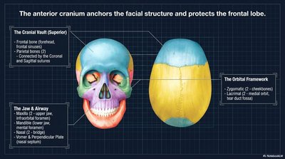
Cranial Structure and Protection
Anterior Cranium and Facial Anchoring
The anterior cranium provides structural support and protection for the brain and facial features:
Cranial Vault: Frontal bone, parietal bones, occipital bone (connected by coronal and sagittal sutures).
Orbital Framework: Zygomatic, lacrimal, and orbital plates.
Jaw & Airway: Mandible, maxilla, vomer, perpendicular plate (nasal septum).
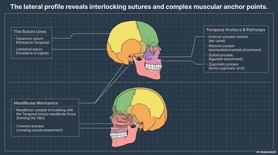
Lateral Skull Profile
Sutures and Muscular Anchor Points
The lateral view of the skull reveals interlocking sutures and attachment sites for muscles:
Suture Lines: Squamous, lambdoid, and coronal sutures.
Temporal Anchors & Pathways: External acoustic meatus, mastoid process, styloid process, zygomatic process, and temporal fossa.
Mandibular Mechanics: Mandibular condyle (TMJ), coronoid process (chewing muscle attachment).
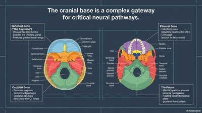
Cranial Base
Neural Pathways and Bone Landmarks
The cranial base is a complex structure that allows passage of critical neural and vascular structures:
Sphenoid Bone: Houses the sella turcica (pituitary gland), features greater/lesser wings.
Ethmoid Bone: Cribriform plate (olfactory foramina), orbital plates.
Occipital Bone: Foramen magnum (spinal cord passage), occipital condyles (articulation with C1 Atlas).
The Palate: Maxilla, palatine process, horizontal plate.
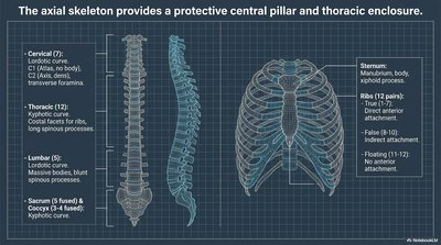
Axial Skeleton
Central Pillar and Thoracic Enclosure
The axial skeleton forms the central support structure and protects vital organs:
Vertebral Column: Cervical (7), thoracic (12), lumbar (5), sacrum (5 fused), coccyx (4 fused).
Thoracic Cage: Sternum (manubrium, body, xiphoid process), ribs (true, false, floating).
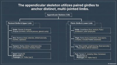
Appendicular Skeleton
Paired Girdles and Limbs
The appendicular skeleton consists of the limbs and their supporting girdles:
Pectoral Girdle & Upper Limb: Clavicle, scapula, humerus, radius, ulna, carpals, metacarpals, phalanges.
Pelvic Girdle & Lower Limb: Coxal bone (ilium, ischium, pubis), femur, tibia, fibula, tarsals, metatarsals, phalanges.
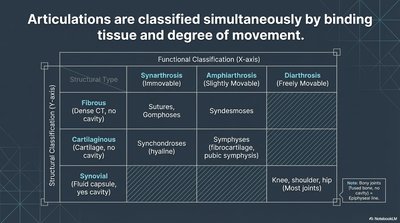
Joint Classification
Binding Tissue and Movement Degree
Joints (articulations) are classified by the tissue binding the bones and their range of movement:
Structural Type | Synarthrosis (Immovable) | Amphiarthrosis (Slightly Movable) | Diarthrosis (Freely Movable) |
|---|---|---|---|
Fibrous | Sutures, gomphoses | Syndesmoses | |
Cartilaginous | Synchondroses (hyaline) | Symphyses (fibrocartilage, pubic symphysis) | |
Synovial | Knee, shoulder, hip (most joints) |
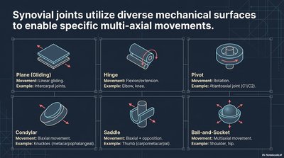
Synovial Joint Types
Mechanical Surfaces and Movements
Synovial joints are categorized by the shape of their articulating surfaces and the movements they allow:
Plane (Gliding): Linear gliding (intercarpal joints).
Hinge: Flexion/extension (elbow, knee).
Pivot: Rotation (atlantoaxial joint).
Condylar: Biaxial movement (knuckles).
Saddle: Biaxial + opposition (thumb).
Ball-and-Socket: Multiaxial movement (shoulder, hip).
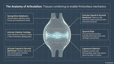
Anatomy of Articulation
Tissues Enabling Frictionless Movement
Several tissues combine to form a functional synovial joint:
Spongy Bone (Epiphysis): Absorbs shock and mechanical stress.
Articular (Hyaline) Cartilage: Provides a smooth, frictionless surface.
Articular Capsule & Synovial Membrane: Encloses the joint, secretes synovial fluid.
Synovial Fluid: Lubricates and nourishes cartilage.
Ligaments & Menisci: Stabilize and restrict abnormal movement.
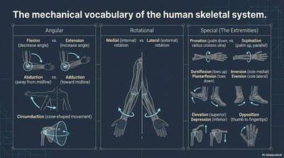
Movements of the Skeletal System
Mechanical Vocabulary
Movements at joints are described using specific anatomical terms:
Angular Movements:
Flexion (decrease angle)
Extension (increase angle)
Abduction (away from midline)
Adduction (toward midline)
Circumduction (cone-shaped movement)
Rotational Movements:
Medial (internal) rotation
Lateral (external) rotation
Special Movements:
Pronation/Supination (forearm)
Dorsiflexion/Plantarflexion (ankle)
Inversion/Eversion (foot)
Elevation/Depression (shoulder)
Opposition (thumb to fingertips)