Back
BackLecture 15 - Impulse Transmission II: Postsynaptic Potentials, Graded vs. Action Potentials, Conduction Velocity, and Neurotransmitters
Study Guide - Smart Notes
 Tailored notes based on your materials, expanded with key definitions, examples, and context.
Impulse Transmission II
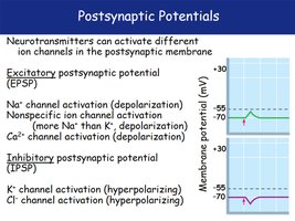
Postsynaptic Potentials
Postsynaptic potentials are changes in membrane potential of the postsynaptic neuron, triggered by neurotransmitter binding to ion channels. These potentials can be either excitatory or inhibitory, depending on the type of ion channel activated.
Excitatory Postsynaptic Potential (EPSP): Caused by activation of Na+ or Ca2+ channels, leading to depolarization.
Inhibitory Postsynaptic Potential (IPSP): Caused by activation of K+ or Cl- channels, leading to hyperpolarization.
Key Point: EPSPs make the neuron more likely to fire an action potential, while IPSPs make it less likely.
Example: Activation of Na+ channels increases membrane potential toward threshold, while Cl- channel activation decreases it.
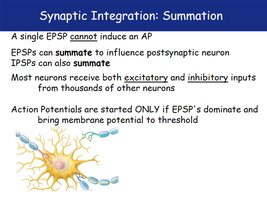
Synaptic Integration: Summation
Neurons integrate multiple postsynaptic potentials through summation. A single EPSP is insufficient to trigger an action potential, but multiple EPSPs or IPSPs can combine to influence the postsynaptic neuron.
Summation: EPSPs and IPSPs can add together (summate) to determine whether the neuron reaches threshold for an action potential.
Excitatory and Inhibitory Inputs: Most neurons receive both types of input from thousands of other neurons.
Threshold: Action potentials occur only if EPSPs dominate and bring the membrane potential to threshold.
Example: A neuron receiving simultaneous EPSPs and IPSPs will only fire if the net effect is depolarizing.
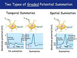
Two Types of Graded Potential Summation
Summation of graded potentials occurs in two main forms: temporal and spatial. Both mechanisms are essential for neural integration and action potential initiation.
Temporal Summation: Multiple EPSPs from a single presynaptic neuron occur in rapid succession, adding together.
Spatial Summation: EPSPs from multiple presynaptic neurons occur simultaneously, combining their effects.
Example: Temporal summation is like repeated taps on a doorbell; spatial summation is like several people pressing the doorbell at once.
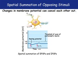
Spatial Summation of Opposing Stimuli
Spatial summation can involve both excitatory and inhibitory inputs. When these occur together, their effects can cancel each other out, preventing the neuron from reaching threshold.
Key Point: The net change in membrane potential depends on the balance of EPSPs and IPSPs.
Example: If an EPSP and an IPSP occur at the same time, the resulting membrane potential may remain unchanged.
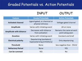
Graded Potentials vs. Action Potentials
Comparison of Graded and Action Potentials
Graded potentials and action potentials are two types of electrical signals in neurons. They differ in their properties, mechanisms, and functions.
Graded Potentials: Local changes in membrane potential, vary in amplitude, can be depolarizing or hyperpolarizing, and decrease with distance.
Action Potentials: All-or-none signals, constant amplitude, only depolarizing, propagate without decrement, and require a threshold.
Refractory Period: Action potentials have a refractory period; graded potentials do not.
Summation: Graded potentials can be summed; action potentials cannot.
Feature | Graded Potentials | Action Potentials |
|---|---|---|
Activated channel | Ligand-gated, chemical or physical stimulus | Voltage-gated channel |
Amplitude | Varies with initiating event | All-or-none |
Amplitude with distance | Decreases with distance from stimulation | Remains the same at each point along axon |
Duration | Varies with initiating event | Constant and brief |
Electrical polarity | Depolarizing (excitatory) or hyperpolarizing (inhibitory) | Only depolarizing |
Threshold | None | Less negative than -55mV |
Refractory Period | None | Yes |
Can be summed | Yes | No |
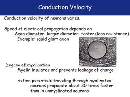
Conduction Velocity
Factors Affecting Conduction Velocity
Conduction velocity refers to the speed at which an action potential travels along a neuron. It is influenced by axon diameter and degree of myelination.
Axon Diameter: Larger diameter axons conduct faster due to lower resistance (e.g., squid giant axon).
Degree of Myelination: Myelin insulates axons, preventing charge leakage and increasing conduction speed.
Example: Myelinated neurons conduct action potentials about 30 times faster than unmyelinated neurons.
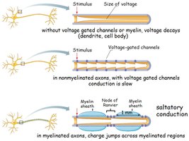
Mechanisms of Conduction
Voltage propagation differs in dendrites, nonmyelinated axons, and myelinated axons. Myelination enables saltatory conduction, where the action potential jumps between nodes of Ranvier.
Without voltage-gated channels or myelin: Voltage decays rapidly.
Nonmyelinated axons: Conduction is slow, as voltage-gated channels are distributed along the axon.
Myelinated axons: Saltatory conduction allows rapid transmission as charge jumps across myelinated regions.
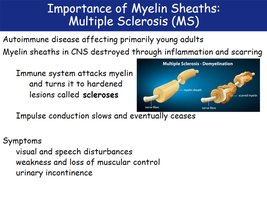
Importance of Myelin Sheaths: Multiple Sclerosis (MS)
Myelin sheaths are critical for fast impulse conduction. Multiple Sclerosis (MS) is an autoimmune disease where myelin in the CNS is destroyed, leading to impaired neural transmission.
Pathology: Immune system attacks myelin, causing hardened lesions (scleroses).
Symptoms: Visual and speech disturbances, muscle weakness, loss of control, urinary incontinence.
Effect: Impulse conduction slows and eventually ceases.
Neurotransmitters and Receptors
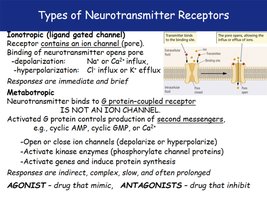
Types of Neurotransmitter Receptors
Neurotransmitter receptors are classified as ionotropic or metabotropic, each with distinct mechanisms and effects.
Ionotropic (Ligand-gated channel): Receptor contains an ion channel; neurotransmitter binding opens the channel for immediate and brief responses.
Metabotropic: Receptor is a G protein-coupled receptor; neurotransmitter binding activates second messengers for indirect, complex, and prolonged responses.
Agonists: Drugs that mimic neurotransmitter action.
Antagonists: Drugs that inhibit neurotransmitter action.
Acetylcholine Receptors (Cholinergic)
Acetylcholine acts on two main types of cholinergic receptors: nicotinic and muscarinic, each with unique properties and locations.
Nicotinic Receptors: Ionotropic, cause Na+ influx and depolarization; found in skeletal muscle, autonomic nervous system, and some CNS pathways.
Muscarinic Receptors: Metabotropic (G-protein coupled), can be excitatory or inhibitory; found in heart, smooth muscle, and glands.
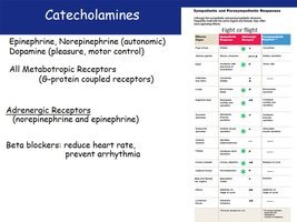
Catecholamines
Catecholamines include epinephrine, norepinephrine, and dopamine, which act through metabotropic receptors and play roles in autonomic function and motor control.
Epinephrine and Norepinephrine: Involved in autonomic responses; act on adrenergic receptors.
Dopamine: Associated with pleasure and motor control.
Beta Blockers: Reduce heart rate and prevent arrhythmia by blocking adrenergic receptors.
Sympathetic Response | Parasympathetic Response |
|---|---|
Fight or flight | Rest and digest |
Increased heart rate | Decreased heart rate |
Bronchodilation | Bronchoconstriction |
Pupil dilation | Pupil constriction |
Reduced digestion | Enhanced digestion |
Beta blockers inhibit sympathetic effects | --- |
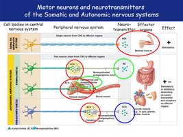
Motor Neurons and Neurotransmitters of the Somatic and Autonomic Nervous Systems
Motor neurons use different neurotransmitters and receptors depending on whether they are part of the somatic or autonomic nervous system.
Somatic Nervous System: Uses acetylcholine (ACh) at the neuromuscular junction to stimulate skeletal muscle.
Autonomic Nervous System: Uses ACh and norepinephrine to regulate smooth muscle, cardiac muscle, and glands.
Effect: Neurotransmitter and receptor type determine whether the response is stimulatory or inhibitory.
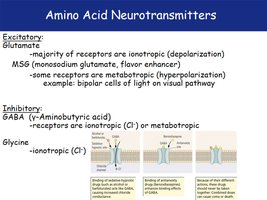
Amino Acid Neurotransmitters
Amino acid neurotransmitters include glutamate (excitatory), GABA, and glycine (inhibitory). Their effects depend on the type of receptor activated.
Glutamate: Major excitatory neurotransmitter; most receptors are ionotropic (depolarizing).
GABA (γ-Aminobutyric acid): Major inhibitory neurotransmitter; receptors can be ionotropic (Cl-) or metabotropic.
Glycine: Inhibitory; ionotropic (Cl-).
Example: MSG (monosodium glutamate) enhances flavor by activating glutamate receptors.