Back
BackInfectious Diseases Affecting the Gastrointestinal Tract: Anatomy & Physiology Study Guide
Study Guide - Smart Notes
 Tailored notes based on your materials, expanded with key definitions, examples, and context.
Gastrointestinal Tract Anatomy and Defenses
Anatomical Features of the Gastrointestinal Tract
The gastrointestinal (GI) tract is a continuous tube extending from the mouth to the anus, responsible for digestion and absorption of nutrients. It consists of eight main sections and four accessory organs that contribute digestive fluids and enzymes.
Main Sections: Mouth, pharynx, esophagus, stomach, small intestine, large intestine, rectum, anus
Accessory Organs: Salivary glands, liver, gallbladder, pancreas
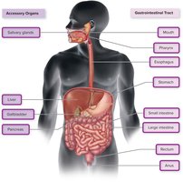
Additional info: The liver is a large visceral organ located in the right abdomen, the stomach is a sac, and the intestines are long and coiled, ending with the rectum and anus.
Natural Defenses of the Gastrointestinal Tract
The GI tract is exposed to a vast number of microorganisms daily, necessitating robust defenses against infection.
Mucus Layer: Coats all intestinal surfaces, providing a physical barrier.
Secretory IgA: Immunoglobulin found on most surfaces, neutralizes pathogens.
Peristalsis: Muscular contractions move food and microorganisms through the tract.
Saliva: Contains lysozyme and lactoferrin, both antimicrobial.
Stomach Fluid: High acidity acts as an antimicrobial agent.
Bile: Also possesses antimicrobial properties.
Gut-associated lymphoid tissue (GALT): Includes tonsils, adenoids, Peyer's patches, and appendix; produces IgA and other immune functions.
Commensal Organisms: Especially abundant in the large intestine, help avoid immune destruction by cloaking themselves with host sugars.
Normal Biota of the Gastrointestinal Tract
Types and Functions of Normal Biota
The GI tract hosts a diverse community of microorganisms, which play essential roles in health and disease.
Oral Cavity: Over 600 species, including Streptococcus, Lactobacillus, Neisseria, Prevotella, Treponema, Veillonella, and Candida albicans (fungus).
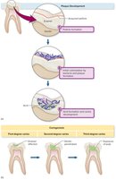
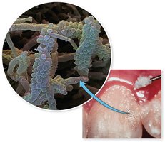
Dental Plaque: Biofilm of bacteria, with alpha-hemolytic streptococci as first colonizers.
Stomach: Contains species such as Bacillus, Clostridium, Staphylococcus, and Streptococcus.
Large Intestine: Billions of microbes per gram; includes Bacteroides, Bifidobacterium, Clostridium, Enterobacter, Escherichia, Fusobacterium, Lactobacillus, Peptostreptococcus, Staphylococcus, Streptococcus, Candida (fungus), protozoa, and archaea.
Functions:
Protective function against pathogens
Assist in digestion and nutrient synthesis (e.g., E. coli synthesizes vitamin K)
Maintain epithelial cell structure
Educate the immune system
Influence host metabolism and disease risk
Gastrointestinal Diseases Caused by Microorganisms
Overview of Foodborne Diseases
Foodborne diseases are a major public health concern, with various pathogens causing illness and death.
Top Causes of Foodborne Illness: Norovirus, Salmonella, Campylobacter, Clostridium, Staphylococcus
Top Causes of Death: Salmonella, Toxoplasma, Listeria, Norovirus, Campylobacter
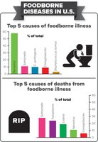
Acute Diarrhea: Highlight Disease
Acute diarrhea is defined as three or more loose stools in a 24-hour period. It is a common condition, especially in children, and can be caused by various pathogens.
Often accompanied by fever, abdominal pain, cramping, nausea, vomiting, and dehydration
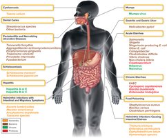
Major causes include Salmonella, Shigella, Shiga-toxin-producing E. coli (STEC), Campylobacter, Clostridioides difficile, Vibrio cholerae, protozoa, and viruses
Salmonella
Salmonella enterica subspecies enterica is the primary cause of salmonellosis, a foodborne illness.
Characteristics: Motile, ferments glucose, produces hydrogen sulfide, resistant to bile and dyes
Transmission: Contaminated animal products (meat, milk, eggs)
Signs and Symptoms: Vomiting, diarrhea, mucosal irritation, blood in stool, fever, septicemia in severe cases
Virulence Factors: Adherence to gut mucosa, endotoxin production
Treatment: Fluid and electrolyte replacement; antibiotics (ciprofloxacin) for severe cases
Shigella
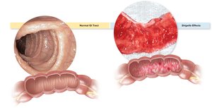
Shigella species cause shigellosis, characterized by frequent, watery stools, fever, intense abdominal pain, and dysentery (bloody diarrhea).
Characteristics: Gram-negative rods, nonmotile, human parasites
Pathogenesis: Invades villus cells of the large intestine, triggers inflammation, releases endotoxin and Shiga toxin
Transmission: Oral route, direct person-to-person contact, low infectious dose
Prevention: Good food hygiene
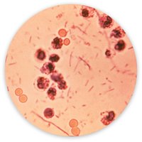
Clostridioides difficile
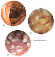
Clostridioides difficile is a gram-positive, endospore-forming rod that causes pseudomembranous colitis, often following broad-spectrum antibiotic therapy.
Pathogenesis: Enterotoxins A and B cause necrosis in the intestinal wall
Signs and Symptoms: Abdominal cramps, fever, leukocytosis, pseudomembrane formation, possible perforation and death
Treatment: Withdraw offending antibiotic, treat with metronidazole or vancomycin
Prevention: Isolation and strict infection control in healthcare settings
Cryptosporidium
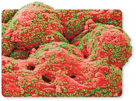
Cryptosporidium is an intestinal protozoan causing gastroenteritis, especially in immunocompromised patients.
Transmission: Ingestion of oocysts in contaminated water or food
Pathogenesis: Oocysts excyst in the intestine, sporozoites attach to epithelium, live intracellularly
Diagnosis: ELISA or acid-fast staining of fecal samples
Treatment: Usually supportive; nitazoxanide for immunocompromised
Giardia lamblia
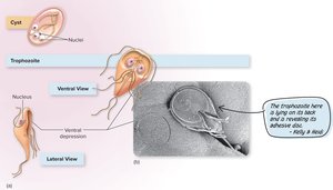
Giardia duodenalis is a flagellated protozoan causing chronic diarrhea, abdominal pain, and flatulence.
Transmission: Ingestion of cysts in contaminated water or food
Pathogenesis: Trophozoites adhere to intestinal wall via ventral disc
Diagnosis: Microscopy of stool samples
Treatment: Tinidazole or nitazoxanide
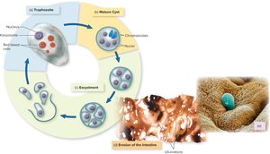
Entamoeba histolytica
Entamoeba histolytica causes amoebiasis, with both intestinal and extraintestinal manifestations.
Pathogenesis: Trophozoites invade mucosa, cause ulcerations and dysentery; can migrate to liver and other organs
Transmission: Ingestion of cysts in contaminated food or water
Treatment: Metronidazole or paromomycin; supportive therapy for fluid/electrolyte replacement
Tooth and Gum Infections
Dental Caries (Tooth Decay)
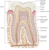
Dental caries is the most common infectious disease in humans, resulting from bacterial metabolism of carbohydrates and acid production.
Causative Agents: Streptococcus mutans and other oral streptococci
Pathogenesis: Bacteria produce sticky polymers (fructans, glucans) and acid, leading to enamel demineralization
Prevention: Dietary restriction of sucrose, regular brushing/flossing, fluoride supplementation
Treatment: Removal and restoration of affected tooth structure
Periodontitis
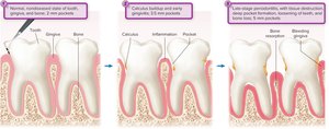
Periodontitis is a progressive disease of the gums and supporting structures of the teeth, often following gingivitis.
Signs and Symptoms: Swelling, redness, bleeding, pocket formation, bone resorption, tooth loosening
Pathogenesis: Polymicrobial biofilms, mineralized plaque (calculus), inflammatory response
Treatment: Removal of calculus/plaque, oral hygiene, surgery, antibiotics
Mumps, Gastritis, and Hepatitis
Mumps
Mumps is a viral infection primarily affecting the parotid salivary glands, causing swelling and systemic symptoms.
Signs and Symptoms: Fever, nasal discharge, muscle pain, malaise, parotitis (cheek swelling)
Transmission: Salivary and respiratory secretions
Prevention: MMR vaccine

Gastritis and Gastric Ulcers
Caused by Helicobacter pylori, which colonizes the stomach mucosa and produces urease to neutralize acidity.
Signs and Symptoms: Burning abdominal pain, ulcers, bloody stools, vomiting
Pathogenesis: Bores through mucus layer, attaches to cells, produces urease
Treatment: Antibiotics and acid suppressors
Hepatitis
Hepatitis is an inflammatory disease of the liver, caused by various viruses (HAV, HBV, HCV, HDV, HEV) and other factors.
Signs and Symptoms: Jaundice, liver swelling, malaise, fever, abdominal discomfort
Transmission: Fecal-oral (HAV, HEV), blood/sexual contact (HBV, HCV, HDV)
Prevention: Vaccines for HAV and HBV; hygiene and safe practices
Treatment: Supportive care, antiviral drugs for chronic cases
Helminthic Infections of the Gastrointestinal Tract
Helminth Diversity and Life Cycles
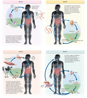
Helminths are parasitic worms classified as nematodes (roundworms), trematodes (flukes), and cestodes (tapeworms). They follow four basic life and transmission cycles.
Cycle A: Eggs ingested, hatch in intestine
Cycle B: Larvae hatch in environment, infect host
Cycle C: Intermediate host (animal), humans ingest infected meat
Cycle D: Complex cycle involving multiple hosts
Cysticercosis (Taenia solium)
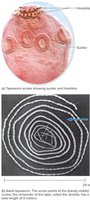
Cysticercosis is caused by the pork tapeworm Taenia solium, with humans as accidental hosts.
Transmission: Ingestion of eggs or cysticerci in undercooked pork
Pathogenesis: Larvae encyst in muscles or brain (neurocysticercosis)
Diagnosis: Imaging, serology
Treatment: Antihelminthic drugs, surgery for severe cases
Schistosomiasis
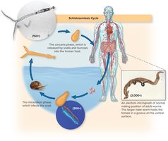
Schistosomiasis is caused by blood flukes (Schistosoma species), with a complex life cycle involving snails as intermediate hosts.
Transmission: Skin penetration by cercariae in contaminated water
Pathogenesis: Adult flukes live in blood vessels, cause liver and bladder disease
Diagnosis: Detection of eggs in stool or urine, serology
Treatment: Antihelminthic drugs
Summary of Infectious Diseases Affecting the GI Tract
The GI tract is susceptible to a wide range of infectious diseases caused by bacteria, viruses, protozoa, and helminths. Understanding the anatomy, normal biota, and defense mechanisms is essential for recognizing and managing these conditions.