Back
BackInfectious Diseases Affecting the Respiratory System: Anatomy, Defenses, and Pathology
Study Guide - Smart Notes
 Tailored notes based on your materials, expanded with key definitions, examples, and context.
Respiratory System Anatomy and Defenses
Overview of the Respiratory Tract
The respiratory tract is divided into upper and lower sections, each with distinct anatomical features and physiological roles. Understanding these divisions is essential for recognizing how infectious diseases affect the respiratory system.
Upper respiratory tract: Includes the mouth, nose, nasal cavity, sinuses, throat (pharynx), epiglottis, and larynx.
Lower respiratory tract: Comprises the trachea, bronchi, bronchioles, and alveoli.
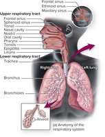
Additional info: The alveoli are the primary sites of gas exchange in the lungs.
Natural Defenses of the Respiratory Tract
The respiratory tract is equipped with multiple defense mechanisms to prevent infection by pathogens.
Nasal hair: Traps large particles and pathogens.
Cilia: Propel trapped particles and microorganisms upward and out of the respiratory tract.
Mucus: Acts as a sticky barrier, trapping invading microorganisms.
Immune defenses: Include complement proteins, antimicrobial peptides, cytokines, macrophages, and secretory IgA.
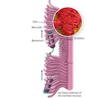
Additional info: The coordinated movement of cilia is known as the mucociliary escalator, which is crucial for clearing pathogens from the airway.
Normal Biota of the Respiratory Tract
Composition and Role of Normal Biota
The respiratory tract harbors a diverse community of commensal microorganisms, which play a protective role by competing with pathogens for resources and space (microbial antagonism).
Common normal biota: Streptococcus pyogenes, Haemophilus influenzae, Streptococcus pneumoniae, Neisseria meningitidis, Staphylococcus aureus, and fungi such as Candida albicans.
Microbial antagonism: For example, Lactobacillus sakei suppresses Corynebacterium tuberculostearicum in the sinuses.
Variation: The composition of the lung microbiome differs in patients with chronic diseases (COPD, asthma, cystic fibrosis) and between smokers and nonsmokers.
Additional info: Disruption of normal biota can predispose individuals to respiratory infections.
Upper Respiratory Tract Diseases
Pharyngitis
Pharyngitis is inflammation of the throat, often presenting with pain, redness, and swelling. Bacterial pharyngitis is typically more severe than viral forms and may be accompanied by fever, headache, and nausea.
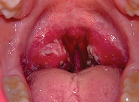
Major causative agents: Viruses (common cold viruses), Streptococcus pyogenes (Group A Streptococcus), and Fusobacterium necrophorum.
Complications: Untreated S. pyogenes infections can lead to scarlet fever (erythrogenic toxin-mediated rash and high fever) and rheumatic fever (autoimmune reaction affecting the heart).
Streptococcus pyogenes: Structure and Virulence
Streptococcus pyogenes is a gram-positive coccus that forms chains and possesses several virulence factors that enhance its pathogenicity.
Structural features: Thick peptidoglycan layer, M-protein fimbriae, protein antigens, hyaluronic acid capsule, and lipoteichoic acid.
Virulence factors: Surface antigens mimic host proteins, superantigen production, resistance to lysozyme, adherence to epithelial cells, and resistance to phagocytosis.
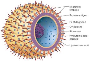
Additional info: The M protein is a key factor in immune evasion and adherence.
Diagnosis of Streptococcal Pharyngitis
Diagnosis involves rapid antigen detection tests and culture of pharyngeal swabs on sheep blood agar, where S. pyogenes produces beta-hemolysis.
Rapid tests: Use antibodies to detect Group A streptococci.
Culture: Beta-hemolysis distinguishes S. pyogenes from other streptococci.
Bacitracin sensitivity: Used to differentiate Group A from other beta-hemolytic streptococci.

Other Upper Respiratory Tract Diseases
The Common Cold: Caused by over 200 viruses (rhinoviruses, coronaviruses, adenoviruses, RSV). Symptoms include sneezing, scratchy throat, and runny nose.
Sinusitis: Can be viral, bacterial, or fungal. Noninfectious causes include allergies and structural abnormalities.
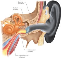
Acute Otitis Media: Middle ear infection often following viral upper respiratory infection. Common agents: Streptococcus pneumoniae, Candida auris. Prevention includes vaccines like Prevnar; treatment may involve antibiotics or tympanic membrane tubes.
Lower Respiratory Tract Diseases
Pneumonia
Pneumonia is an inflammatory condition of the lung in which fluid fills the alveoli. It can be caused by bacteria, viruses, or fungi, and is a leading cause of death in children under five worldwide.
Community-acquired pneumonia: Common agents include Streptococcus pneumoniae, Legionella pneumophila, Mycoplasma pneumoniae, Chlamydophila pneumoniae, Histoplasma capsulatum, and hantavirus.
Healthcare-associated pneumonia: Often associated with mechanical ventilation and caused by MRSA, Klebsiella pneumoniae, Pseudomonas aeruginosa, and others.
Additional info: Vaccination (e.g., PPSV23, PCV13) is recommended for older adults to prevent pneumococcal pneumonia.
Influenza
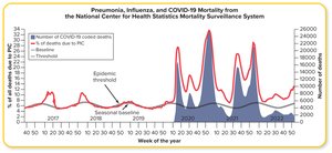
Influenza is a viral infection with seasonal epidemics and occasional pandemics. It is caused by influenza A, B, and C viruses, which are known for their genetic variability.
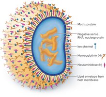
Structure: Spherical particles with a lipoprotein envelope, hemagglutinin (H), neuraminidase (N), and ion channels. The genome consists of 8 RNA segments.
Antigenic drift: Gradual mutation of H and N antigens, requiring annual vaccine updates.
Antigenic shift: Major genetic reassortment between different influenza viruses, often resulting in pandemics.
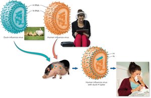
Additional info: Antigenic shift can produce novel strains to which the population has little immunity.
Diagnosis and Epidemiology of Influenza
Diagnosis is primarily by RT-PCR. Influenza spreads via aerosols, droplets, and fomites, with higher transmission in crowded and poorly ventilated environments.
Other Lower Respiratory Tract Diseases
Pertussis (Whooping Cough): Caused by Bordetella pertussis. Prevented by DTaP vaccine.
Respiratory Syncytial Virus (RSV): Major cause of lower respiratory tract infections in infants and young children. Prevention includes passive antibody for high-risk children and vaccines for older adults.
Tuberculosis
Overview and Pathogenesis
Tuberculosis (TB) is a chronic infectious disease caused by Mycobacterium tuberculosis. It primarily affects the lungs but can disseminate to other organs.
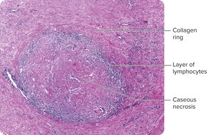
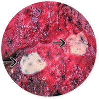
Primary TB: Bacteria multiply in macrophages, leading to tubercle formation and caseous necrosis.
Secondary (Reactivation) TB: Dormant bacteria reactivate, causing severe symptoms and lung damage.
Extrapulmonary TB: Involvement of lymph nodes, kidneys, bones, genital tract, or brain, often fatal in immunosuppressed patients.
Diagnosis, Prevention, and Treatment of Tuberculosis
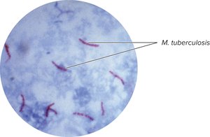
Diagnosis: Tuberculin skin test (Mantoux), interferon-gamma release assays, gene amplification, and acid-fast staining.
Prevention: Limiting exposure, patient isolation, and BCG vaccine (not used in the US).
Treatment: Long-term antibiotic regimens (4-9 months). Noncompliance leads to multidrug-resistant TB (MDR-TB) and extensively drug-resistant TB (XDR-TB).
Additional info: MDR-TB is resistant to at least isoniazid and rifampin; XDR-TB is resistant to additional drugs and has a high mortality rate.
Summary Table: Major Infectious Diseases of the Respiratory System
Disease | Causative Agent(s) | Transmission | Key Features | Prevention/Treatment |
|---|---|---|---|---|
Pharyngitis | Streptococcus pyogenes, viruses, Fusobacterium necrophorum | Droplets, direct contact | Sore throat, fever, risk of rheumatic fever | Hand hygiene, antibiotics (penicillin) |
Pneumonia | Streptococcus pneumoniae, viruses, fungi | Droplets, aspiration | Alveolar inflammation, deadly in children | Vaccines, antibiotics, supportive care |
Influenza | Influenza A, B, C viruses | Droplets, fomites | Fever, cough, antigenic drift/shift | Annual vaccine, antivirals |
Tuberculosis | Mycobacterium tuberculosis | Airborne droplets | Chronic cough, caseous necrosis | Long-term antibiotics, BCG vaccine |