 Back
Innate and Adaptive Immunity: The Immune System’s Defenses
Study Guide - Smart Notes
 Tailored notes based on your materials, expanded with key definitions, examples, and context.
Introduction to Immunity
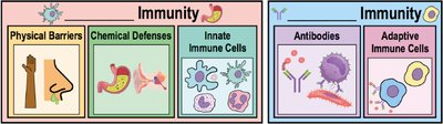
The immune system protects the body from disease-causing microbes and environmental threats through a complex network of defenses. Immunity is the ability to eliminate pathogens and protect against harmful substances, while susceptibility is the lack of immunity to a particular threat. There are two main types of immunity: innate (non-specific) and adaptive (specific). Innate immunity provides immediate, broad protection, while adaptive immunity develops over time and targets specific pathogens.
Overview of Immune Defenses
Innate Immunity: Present at birth, provides rapid, non-specific defense against a wide range of pathogens.
Adaptive Immunity: Develops after exposure to specific antigens, providing targeted and long-lasting protection.
Antigens and Antibodies
Key Concepts
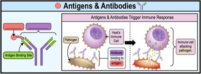
Antigen: Any toxin or foreign substance that induces an immune response, specifically the production of antibodies.
Antibody: Y-shaped protein produced by B cells that recognizes and binds to specific antigens, marking them for destruction by immune cells.
Antigens and antibodies are central to adaptive immunity. When antibodies bind to antigens, they trigger immune responses that help eliminate pathogens.
Lines of Defense in Innate Immunity
First-Line Defenses
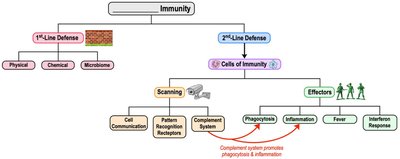
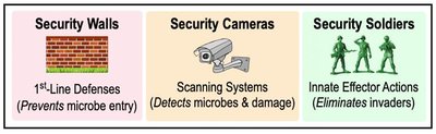
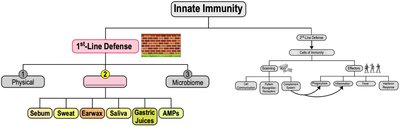
The first line of defense consists of physical, chemical, and microbiome barriers that prevent pathogens from entering the body.
Physical Barriers: Skin and mucous membranes form protective layers.
Chemical Barriers: Bodily fluids (e.g., saliva, stomach acid), antimicrobial peptides, and secretions like sebum and sweat inhibit microbial growth.
Microbiome: Normal microbiota compete with pathogens for resources and space.
Second-Line Defenses
If pathogens bypass the first line, the second line of defense involves immune cells and processes that detect and eliminate invaders.
Scanning Systems: Detect signs of infection (e.g., pattern recognition receptors).
Innate Effector Actions: Include phagocytosis, inflammation, fever, and interferon response.
Physical Barriers
Skin
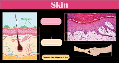
The skin is a multi-layered barrier composed of tightly packed epithelial cells. The outermost layer contains keratin, a water-repelling protein that inhibits microbial growth. Shedding of dead skin cells removes attached microbes. Beneath the epidermis is the dermis, a tough connective tissue layer, followed by the subcutaneous fat layer.
Mucous Membranes
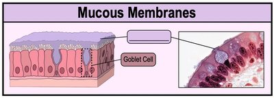
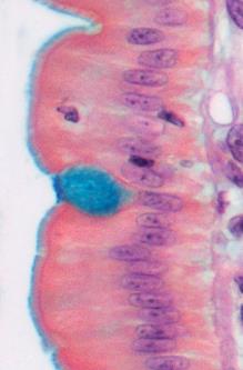
Mucous membranes line the digestive, respiratory, and genitourinary tracts. They consist of epithelial cells and connective tissue that secrete mucus, trapping microbes and preventing tissue dehydration. Specialized mechanisms, such as the mucociliary escalator in the respiratory tract, move trapped microbes out of the body.
Chemical Barriers
Chemical defenses include secretions and substances that inhibit microbial growth:
Sebum: Produced by sebaceous glands, contains fatty acids that lower skin pH and inhibit microbes.
Sweat: Contains lysozyme, an enzyme that degrades bacterial cell walls.
Earwax: Physically blocks entry and maintains acidic pH.
Saliva: Contains enzymes (e.g., lysozyme) that inhibit microbial growth.
Gastric Juice: Highly acidic, destroys most ingested pathogens.
Antimicrobial Peptides (AMPs): Short amino acid chains (e.g., defensins) that disrupt microbial membranes.
Normal Microbiota
The normal microbiota (microbiome) consists of resident microbes that protect against pathogens by occupying attachment sites and producing antimicrobial substances. Disruption of the microbiome (e.g., by antibiotics) can increase susceptibility to infection. Probiotics and prebiotics support a healthy microbiome.
Cells of the Immune System
Hematopoiesis
All blood cells develop from hematopoietic stem cells in the bone marrow. These stem cells differentiate into myeloid and lymphoid progenitor cells, which give rise to red blood cells, platelets, and white blood cells (leukocytes).
Leukocytes
Granulocytes: White blood cells with visible granules (e.g., neutrophils, eosinophils, basophils).
Agranulocytes: White blood cells without visible granules (e.g., monocytes, lymphocytes).
Granulocytes
Neutrophils: Most abundant, first responders, phagocytose and destroy microbes.
Eosinophils: Defend against parasites, involved in allergic reactions.
Basophils: Release histamine during inflammation and allergic responses.
Agranulocytes
Monocytes: Differentiate into macrophages (phagocytic, reside in tissues) and dendritic cells (initiate adaptive immunity).
Lymphocytes: Include B cells, T cells (adaptive immunity), and natural killer (NK) cells (innate immunity).
Cell Communication in Immunity
Immune cells communicate via surface receptors, adhesion molecules, and cytokines:
Surface Receptors: Proteins that detect external signals and trigger cellular responses.
Adhesion Molecules: Enable immune cells to attach to other cells and migrate to infection sites.
Cytokines: Soluble proteins that regulate immune responses (e.g., chemokines, interleukins, interferons, tumor necrosis factor).
Pattern Recognition Receptors (PRRs)
PRRs detect molecular patterns associated with pathogens (MAMPs/PAMPs) or cell damage (DAMPs). Types include:
Toll-Like Receptors (TLRs): Detect microbial molecules on cell surfaces or within endosomes.
C-Type Lectin Receptors (CLRs): Bind carbohydrates on microbial surfaces.
NOD-Like Receptors (NLRs): Detect intracellular MAMPs/DAMPs, trigger inflammasome formation.
RIG-Like Receptors (RLRs): Detect viral RNA in the cytoplasm.
The Complement System
The complement system is a group of inactive proteins in the blood that, when activated, enhance immune responses. Activation occurs via three pathways:
Alternative Pathway: Triggered by C3b binding to microbial surfaces.
Lectin Pathway: Initiated by mannose-binding lectins binding to microbial carbohydrates.
Classical Pathway: Activated by antibodies bound to antigens.
All pathways converge on the activation of C3 convertase, leading to:
Opsonization (C3b coats microbes for phagocytosis)
Cell lysis (membrane attack complex forms pores in microbial membranes)
Inflammatory response (C3a, C5a recruit and activate immune cells)
Phagocytosis
Phagocytosis is the process by which immune cells (e.g., macrophages, neutrophils) engulf and digest pathogens. Steps include:
Chemotaxis: Movement toward infection site.
Recognition & Attachment: Binding to microbes directly or via opsonins.
Engulfment: Formation of a phagosome.
Phagolysosome Formation: Fusion with lysosome.
Destruction & Digestion: Enzymatic breakdown of microbe.
Exocytosis: Expulsion of debris.
Inflammation
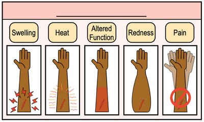
Inflammation is a coordinated response to infection or tissue damage, characterized by swelling, heat, altered function, redness, and pain. Acute inflammation is short-term and resolves quickly, while chronic inflammation is long-term and can lead to tissue damage (e.g., granuloma formation in tuberculosis).
Fever
Fever is an elevated body temperature that inhibits microbial growth and enhances immune responses. It is triggered by pyrogens (endogenous or exogenous) that act on the hypothalamus.
Interferon Response
Interferons (IFNs) are cytokines that provide antiviral protection by signaling neighboring cells to produce antiviral proteins (AVPs). If a cell becomes infected, AVPs are activated, leading to apoptosis and limiting viral spread.
Introduction to Adaptive Immunity
Adaptive immunity develops over time and targets specific pathogens. It consists of two main components:
Cell-Mediated Immunity: T cells target and destroy infected host cells.
Humoral Immunity: B cells produce antibodies that neutralize extracellular pathogens.
Antigens and Antigen Presentation
Adaptive immunity is triggered by antigens. B cells respond to free antigens, while T cells respond to antigens presented by antigen-presenting cells (APCs) on major histocompatibility complex (MHC) molecules.
T Lymphocytes (T Cells)
T Cell Receptors (TCRs): Recognize antigens presented on MHC molecules.
Cytotoxic T Cells (CD8+): Induce apoptosis in infected cells presenting endogenous antigens on MHC I.
Helper T Cells (CD4+): Produce cytokines to activate other immune cells, recognize antigens on MHC II.
Regulatory T Cells (Tregs): Suppress self-reactive T cells and prevent autoimmunity.
B Lymphocytes (B Cells) and Antibodies
B Cell Receptors (BCRs): Recognize free antigens.
Plasma Cells: Effector B cells that secrete antibodies.
Memory B Cells: Provide rapid response upon re-exposure to the same antigen.
Antibody Structure and Classes
Antibodies (immunoglobulins) are Y-shaped proteins with two light and two heavy chains, each with variable and constant regions. There are five main classes:
Class | Main Function |
|---|---|
IgG | Most abundant, crosses placenta, activates complement |
IgA | Protects mucous membranes, found in secretions |
IgM | First produced, activates complement, pentameric |
IgE | Allergic responses, defense against parasites |
IgD | B cell receptor, activation/maturation |
Outcomes of Antibody Binding
Opsonization: Enhances phagocytosis
Neutralization: Blocks pathogen/toxin activity
Agglutination: Clumps pathogens for easier removal
Complement Activation: Triggers classical pathway
Antibody-Dependent Cell-Mediated Cytotoxicity (ADCC): NK cells kill antibody-coated cells
Clonal Selection and Immune Memory
Clonal selection ensures that only B and T cells with receptors specific to an antigen proliferate. Memory cells provide a faster and stronger response upon subsequent exposures (secondary immune response).
Immune Tolerance
Immune tolerance prevents inappropriate responses to harmless or self-antigens. Central and peripheral tolerance mechanisms eliminate or inactivate self-reactive lymphocytes, preventing autoimmune diseases.
Summary Table: Innate vs. Adaptive Immunity
Feature | Innate Immunity | Adaptive Immunity |
|---|---|---|
Specificity | Non-specific | Highly specific |
Response Time | Immediate (hours) | Delayed (days) |
Memory | None | Yes |
Main Components | Barriers, phagocytes, NK cells, complement | B cells, T cells, antibodies |
