 Back
BackInnate Defenses of the Immune System: ANP Study Notes
Study Guide - Smart Notes
 Tailored notes based on your materials, expanded with key definitions, examples, and context.
Immune System Overview
Introduction to the Immune System
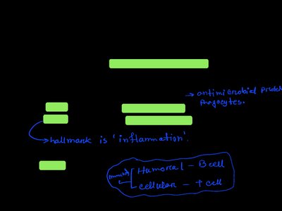
The immune system is a functional system responsible for defending the body against disease-causing agents. It consists of two main intrinsic defense systems: the innate (nonspecific) defense system and the adaptive (specific) defense system. The innate system provides immediate, general protection, while the adaptive system targets specific threats and takes longer to activate.
Innate Defense System: First and second lines of defense; rapid response; non-specific.
Adaptive Defense System: Third line of defense; specific; slower to respond.
Innate (Nonspecific) Defenses
Surface Barriers
Surface barriers are the body's first line of defense, preventing pathogen entry through physical and chemical means.
Physical Barrier: Keratinized epidermis forms a tough, protective layer.
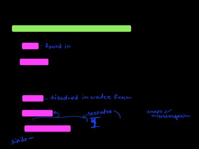
Chemical Barriers:
Acid: Found in skin, vaginal, and stomach secretions; inhibits microbial growth.
Enzymes: Present in saliva, respiratory mucus, lacrimal fluid, and stomach; destroy pathogens.
Mucin: Thick mucus in digestive and respiratory tracts traps microorganisms.
Defensins: Antimicrobial peptides in mucous membranes and skin.
Other Chemicals: Sebum and sweat contain substances toxic to bacteria.
Internal Defenses
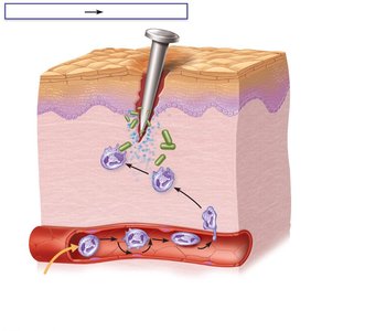
If pathogens bypass surface barriers, the body employs internal defenses, including phagocytes, natural killer cells, inflammation, antimicrobial proteins, and fever.
Phagocytes
Natural Killer (NK) Cells
Inflammation
Antimicrobial Proteins
Fever
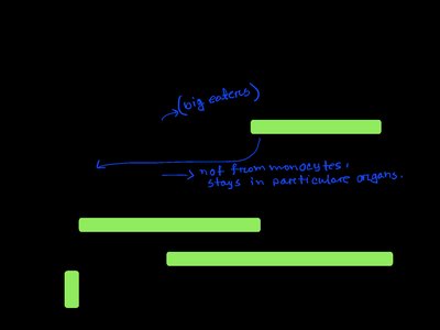
Phagocytes
Phagocytes are cells that engulf and digest pathogens and debris. The main types include:
Macrophages: Develop from monocytes; chief phagocytic cells; can be free or fixed in tissues.
Neutrophils: Most abundant white blood cells; rapidly respond to infection.
Eosinophils: Kill parasitic worms by releasing enzymes; not true phagocytes.
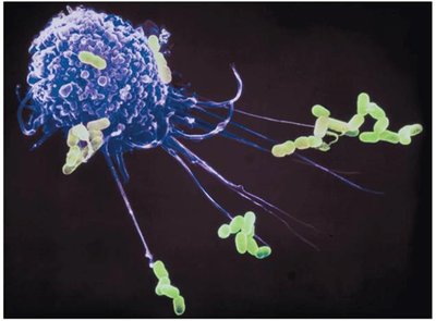
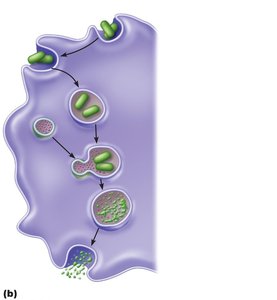
Mechanism of Phagocytosis
Phagocytosis involves several steps:
Phagocyte adheres to pathogens or debris.
Phagocyte forms pseudopods, engulfing particles into a phagosome.
Lysosome fuses with the phagosome, forming a phagolysosome.
Lysosomal enzymes digest the particles.
Exocytosis removes indigestible material.
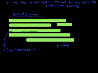
Opsonization
Opsonization is the process by which pathogens are coated with molecules (opsonins) that enhance their recognition and ingestion by phagocytes. Some bacteria evade this by covering their surface markers with a capsule.
Other Mechanisms of Phagocytosis
Respiratory Burst: Helper T cells stimulate phagocytes to produce free radicals and oxidizing chemicals, increasing the destruction of resistant pathogens.
Defensins: Neutrophils release antimicrobial peptides.
Natural Killer (NK) Cells
Natural Killer (NK) cells are large granular lymphocytes that target and destroy virus-infected and cancerous cells before the adaptive immune system is activated. They recognize abnormal cells by the absence of "self" markers and induce apoptosis (programmed cell death).
Non-specific; do not target specific pathogens.
Enhance the inflammatory response.
Inflammatory Response
The inflammatory response is triggered by trauma, heat, chemicals, or infectious agents. It serves to prevent the spread of damaging agents, dispose of debris and pathogens, alert the adaptive immune system, and initiate tissue repair.
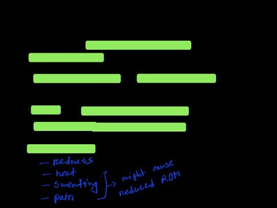
Cardinal Signs of Inflammation: Redness, heat, swelling, pain.
Chemical Mediators of Inflammation
Inflammation is mediated by chemicals such as histamine, kinins, prostaglandins, leukotrienes, and complement proteins. These mediators cause vasodilation (leading to redness and heat) and increased vascular permeability (leading to swelling and pain).
Phagocytic Mobilization
Phagocytes are mobilized to the site of injury in a series of steps:
Leukocytosis: Neutrophils enter blood from bone marrow.
Margination: Neutrophils cling to capillary walls.
Diapedesis: Neutrophils squeeze out of capillaries.
Chemotaxis: Neutrophils follow chemical signals to the site of infection.
Pus and Abscess Formation
Pus consists of dead and dying neutrophils, tissue cells, and pathogens. If not cleared, it may form an abscess, a walled-off sac of pus.
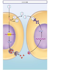
Antimicrobial Proteins
Antimicrobial proteins attack microorganisms directly or inhibit their reproduction. The two most important types are interferons and complement proteins.
Interferons
Interferons (IFNs) are small proteins secreted by virus-infected cells. They enter neighboring cells, inducing the production of antiviral proteins that block viral replication.
Complement System
The complement system consists of over 20 blood proteins that circulate in an inactive state. When activated, they enhance both innate and adaptive defenses by:
Promoting inflammation
Enhancing phagocytosis (opsonization and chemotaxis)
Causing cell lysis via the membrane attack complex (MAC)
There are three pathways for complement activation: classical, lectin, and alternative.
Fever
Fever is an innate defense mechanism in which the body's thermostat is reset upwards in response to pyrogens (substances released by pathogens or immune cells). Moderate fever helps by sequestering iron and zinc in the liver and spleen and increasing metabolic rate, but high fever can be harmful by denaturing enzymes and proteins.
Review Questions
What are the cardinal signs of inflammation and what causes them?
Under what circumstances might NK cells kill our own cells?
What is opsonization?
What distinguishes the innate from the adaptive defense system?
What is the first line of defense against disease?
