Back
BackInnate Immunity: Internal Defenses and the Inflammatory Response
Study Guide - Smart Notes
 Tailored notes based on your materials, expanded with key definitions, examples, and context.
Innate Immunity: Internal Defenses
Overview of Innate Immunity
Innate immunity provides a rapid, non-specific response to pathogens and consists of antimicrobial molecules and specialized cells. It acts as the body's second line of defense after surface barriers are breached.
Phagocytic cells: Macrophages, neutrophils, and eosinophils ingest pathogens and debris.
Nonphagocytic cells: Include natural killer (NK) cells, dendritic cells, and basophils.
Phagocytic Cells
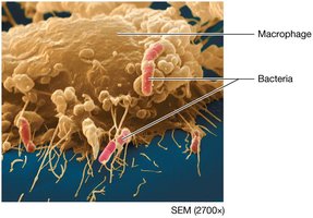
Phagocytes are essential for engulfing and destroying pathogens. They include macrophages, neutrophils, and eosinophils.
Macrophages: Derived from monocytes, these cells reside in tissues and are activated by pathogen molecules, damaged cells, or signals from adaptive immunity. They ingest pathogens and debris, kill with chemicals (e.g., hydrogen peroxide, hypochlorous acid), and present antigens to T cells, initiating adaptive responses.
Neutrophils: Most numerous granulocyte, highly effective at destroying bacteria. They release cytotoxic granules and are recruited to damaged tissues by chemical signals.
Eosinophils: Migrate from blood to tissues, primarily targeting parasitic pathogens by releasing granule contents that damage or destroy parasites.
Antigen-Presenting Cells (APCs)
APCs display pathogen antigens on their membranes, activating T cells and enhancing immune responses.
Macrophages: Present antigens to T cells, which then secrete substances to further activate macrophages.
Dendritic cells: Most important APCs, presenting antigens to T and B cells.
Nonphagocytic Cells
These cells contribute to innate immunity through cytotoxic actions and inflammation.
NK cells: Recognize and destroy cancerous or virus-infected cells, secrete cytokines to activate macrophages.
Basophils and mast cells: Release inflammatory mediators, especially during allergic responses.
Antimicrobial Proteins
Complement System
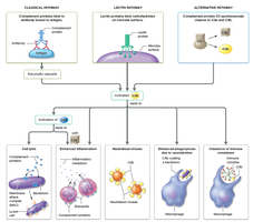
The complement system consists of over 20 plasma proteins produced mainly by the liver. These proteins circulate in inactive forms and are activated by three pathways:
Classical pathway: Initiated by complement proteins binding to antibodies attached to antigens.
Lectin pathway: Triggered by lectins binding to microbial carbohydrates.
Alternative pathway: Activated when complement proteins encounter foreign cells.
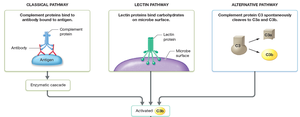
All pathways converge at the activation of C3, which is cleaved to C3b, then C5 to C5b, leading to several immune effects.
Main Effects of Activated Complement Proteins
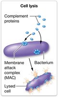
Cell lysis: C5b forms the membrane attack complex (MAC), which creates pores in pathogen membranes, causing lysis.
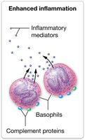
Enhanced inflammation: Complement proteins trigger basophils and mast cells to release inflammatory mediators.
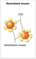
Neutralization of viruses: C3b and MAC components bind to viruses, blocking infection.
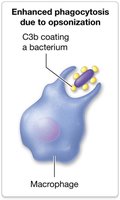
Opsonization: C3b binds to pathogens, enhancing phagocyte binding and phagocytosis.
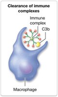
Clearance of immune complexes: C3b binds to antigen-antibody complexes, promoting their removal by phagocytes.
Cytokines
Cytokines are proteins produced by immune cells that regulate and enhance immune responses.
Tumor necrosis factor (TNF): Secreted by macrophages, attracts and activates phagocytes, stimulates cytokine release.
Interferons: Produced in response to viral or intracellular bacterial infection, inhibit viral replication.
Interleukins: Produced by leukocytes, stimulate neutrophil and NK cell production, trigger interferon production, activate T cells.
Additional info: Many cytokines induce flu-like symptoms, including fever, chills, and aches.
The Inflammatory Response
Stages of Inflammation
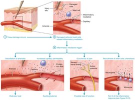
The inflammatory response is triggered by cell damage and consists of two main stages: release of inflammatory mediators and phagocyte arrival.
Stage 1: Damaged cells and mast cells release mediators (histamine, serotonin, cytokines, bradykinin, prostaglandins, leukotrienes). Complement proteins also trigger mediator release.
Cardinal signs: Redness, heat, swelling (edema), and pain.
Vasodilation: Mediators relax arterioles, increasing blood flow and causing redness and heat.
Increased capillary permeability: Protein-rich fluid leaks into tissues, causing swelling.
Pain: Mediators trigger sensory neurons, causing pain and temporary loss of function.
Recruitment: Leukocytes are attracted to the site (chemotaxis).
Phagocyte Response
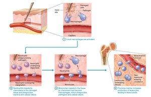
Phagocytes arrive in stages to contain and clear pathogens.
Macrophages: First responders, activated within minutes, phagocytize pathogens and debris.
Neutrophils: Attracted by mediators and complement proteins, leave blood (margination), squeeze into tissue (diapedesis), destroy bacteria and debris.
Monocytes: Migrate to tissue, become macrophages, continue phagocytosis.
Leukocytosis: Cytokines stimulate bone marrow to produce more neutrophils and monocytes, increasing white cell count.
Pus: Accumulation of dead leukocytes, tissue cells, and fluid.
Anti-inflammatory Medications
Mechanisms and Types
Medications reduce inflammation and pain by blocking production of prostaglandins and leukotrienes.
NSAIDs: Inhibit cyclooxygenase, preventing prostaglandin formation (e.g., ibuprofen).
Corticosteroids: Mimic cortisol, inhibit both prostaglandin and leukotriene formation, used for severe or allergy-related inflammation (e.g., cortisone, prednisone).
Additional info: Prostaglandins and leukotrienes are derived from arachidonic acid, cleaved by phospholipase A2.
Fever
Definition and Mechanism
Fever is a body temperature above the normal range (36–38°C or 97–99°F), serving as a warning sign of inflammation.
Initiation: Pyrogens released from damaged cells or bacteria act on the hypothalamus, resetting the body's thermostat.
Response: Hypothalamus triggers shivering and increased muscle activity to raise temperature.
Resolution: When the hypothalamus resets to normal, mechanisms such as sweating and vasodilation lower body temperature.
Additional info: Negative feedback loops regulate body temperature during fever.
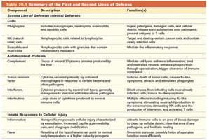
Summary of First and Second Lines of Defense
Table: Surface Barriers and Internal Defenses
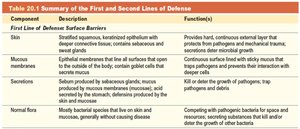
The following tables summarize the components and functions of the body's first and second lines of defense.
Component | Description | Function(s) |
|---|---|---|
Skin | Stratified squamous, keratinized epithelium with sweat and sebaceous glands | Provides hard, continuous external layer that protects from pathogens and mechanical trauma; secretes substances with antimicrobial properties |
Mucous membranes | Epithelium lining all surfaces that open to the outside; contains goblet cells and mucus | Continuous surface lined with sticky mucus that traps pathogens and prevents their interaction with deeper cells |
Secretions | Substances produced by sebaceous glands, mucus, and saliva | Kill or deter the growth of pathogens; trap pathogens and debris |
Normal flora | Bacterial species on skin and mucosa | Compete with pathogenic bacteria for space and resources, reducing their survival and growth |
Component | Description | Function(s) |
|---|---|---|
Phagocytes | Macrophages, neutrophils, eosinophils | Ingest pathogens, damaged cells, and debris |
NK cells | Nonphagocytic lymphocyte-related cells | Target and destroy certain cancer and virus-infected cells |
Basophils and mast cells | Granulocytes | Mediate the inflammatory response |
Complement proteins | Group of 20+ plasma proteins | Mediate cell lysis, enhance inflammation, opsonize pathogens, clear immune complexes |
Tumor necrosis factor | Cytokine secreted by macrophages | Attracts and activates phagocytes, stimulates cytokine release |
Interferons | Cytokines produced by several cell types | Block viral replication, activate immune cells |
Interleukins | Leukocyte-produced cytokines | Stimulate neutrophil and NK cell production, trigger interferon production, activate T cells |
Inflammatory response | Innate response to cell injury | Attracts phagocytes, increases capillary permeability, causes cardinal signs of inflammation |
Fever | Body temperature above normal | Enhances immune response, inhibits pathogen growth |