 Back
BackInnate Immunity: The First and Second Lines of Defense
Study Guide - Smart Notes
 Tailored notes based on your materials, expanded with key definitions, examples, and context.
The Immune System: Overview
Functional Organization of Immunity
The immune system provides resistance to disease through two major intrinsic systems: innate (nonspecific) defenses and adaptive (specific) defenses. These systems are intertwined and collectively protect the body from pathogens and harmful agents.
Innate defenses: Provide immediate, nonspecific protection via surface barriers and internal mechanisms.
Adaptive defenses: Target specific foreign substances and require more time to mount a response.
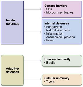
Innate Immunity
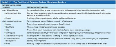
First Line of Defense: Surface Barriers
The first line of defense consists of surface barriers such as skin and mucous membranes, which physically and chemically prevent pathogen entry. These barriers are supported by secretions that inhibit or destroy microorganisms.
Skin: Acts as a mechanical barrier; keratin resists acids, bases, enzymes, and toxins.
Mucous membranes: Provide similar mechanical protection in respiratory, digestive, and urogenital tracts.
Protective chemicals: Include acid mantle, enzymes (e.g., lysozyme), mucin, defensins, lipids in sebum, and dermicidin in sweat.
Respiratory modifications: Mucus-coated hairs and cilia trap and remove pathogens.
CATEGORY/ASSOCIATED ELEMENTS | PROTECTIVE MECHANISM |
|---|---|
Intact skin epidermis | Mechanical barrier preventing entry of pathogens and harmful substances |
Acid mantle of skin | Skin secretions make epidermal surface acidic, inhibiting bacterial growth |
Keratin | Resists acids, alkalis, and bacterial enzymes |
Intact mucous membranes | Mechanical barrier in respiratory, digestive, and urogenital tracts |
Mucus | Traps microorganisms in nasal and digestive passages |
Nasal hairs | Filter and trap microorganisms in nasal passages |
Cilia | Propel debris-laden mucus away from nasal cavity and lower respiratory passages |
Gastric juice | Destroys pathogens with acid and protein-digesting enzymes |
Acid mantle of vagina | Inhibits growth of bacteria and fungi in female reproductive tract |
Lacrimal secretion; saliva | Contains lysozyme, an enzyme that destroys bacteria |
Urine | Acidic pH inhibits bacterial growth; flushes urinary tract |
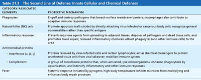
Second Line of Defense: Cells and Chemicals
If pathogens breach surface barriers, the innate system activates internal defenses, including phagocytes, natural killer (NK) cells, inflammation, antimicrobial proteins, and fever. These mechanisms inhibit the spread of invaders and initiate repair processes.
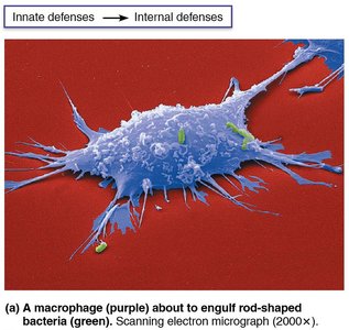
Phagocytes: White blood cells that ingest and digest foreign invaders.
Natural killer (NK) cells: Destroy virus-infected and cancerous cells.
Inflammatory response: Prevents spread, disposes of debris, alerts adaptive immunity, and initiates repair.
Antimicrobial proteins: Interferons and complement proteins attack microorganisms or hinder their reproduction.
Fever: Systemic response that inhibits microbial growth and enhances repair.
CATEGORY/ASSOCIATED ELEMENTS | PROTECTIVE MECHANISM |
|---|---|
Phagocytes | Engulf and destroy pathogens; macrophages contribute to adaptive immune responses |
Natural killer (NK) cells | Induce apoptosis in virus-infected/cancerous cells; recognize general abnormality |
Inflammatory response | Prevents spread, disposes debris/pathogens, alerts adaptive immunity, promotes repair |
Antimicrobial proteins (Interferons, Complement) | Block viral reproduction, enhance phagocytosis, amplify inflammation |
Fever | High temperature inhibits microbes and enhances repair |
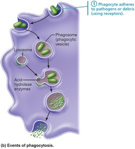
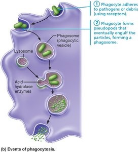
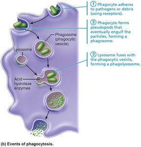
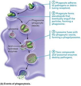
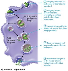
Phagocytosis: Mechanism and Steps
Phagocyte Types and Function
Phagocytes are specialized white blood cells that ingest and digest pathogens. Neutrophils are the most abundant, while macrophages are the most robust and can be free or fixed in tissues.
Steps of Phagocytosis
Phagocytosis is a multi-step process by which phagocytes eliminate pathogens:
Recognition and adherence: Phagocyte binds to pathogen's carbohydrate signature. Opsonization by antibodies or complement proteins enhances this step.
Engulfment: Cytoplasmic extensions (pseudopods) surround the pathogen, forming a phagosome.
Fusion: Phagosome fuses with a lysosome, forming a phagolysosome.
Digestion: Lysosomal enzymes and toxic compounds destroy the pathogen.
Exocytosis: Indigestible material is expelled from the cell.
Inflammation: Tissue Response to Injury
Overview and Benefits
Inflammation is triggered by tissue injury due to trauma, heat, chemicals, or infection. It prevents the spread of damaging agents, disposes of debris, alerts adaptive immunity, and initiates repair.
Cardinal signs: Redness, heat, swelling, pain (sometimes impairment of function).
Stages: Inflammatory chemical release, vasodilation and increased permeability, phagocyte mobilization.
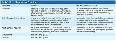
Inflammatory Chemicals
Various chemicals mediate inflammation, including histamine, kinins, prostaglandins, complement, and cytokines. These substances promote vasodilation, increase capillary permeability, and attract phagocytes.
Chemical | Source | Physiological Effects |
|---|---|---|
Histamine | Mast cells, basophils | Vasodilation, increased permeability, attracts phagocytes |
Kinins | Plasma proteins | Same as histamine, plus chemotaxis |
Prostaglandins | Fatty acid molecules | Same as histamine, plus pain |
Complement | Plasma proteins | Enhances inflammation, opsonization |
Cytokines | Various cells | Regulate immune responses |
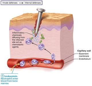
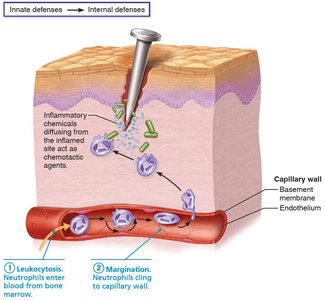
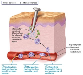
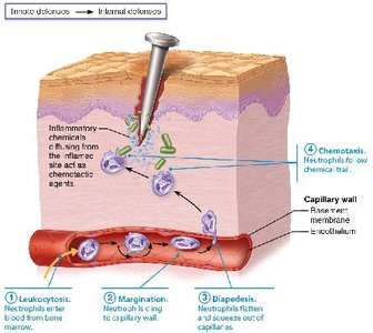
Phagocyte Mobilization
Phagocyte mobilization is a four-step process during inflammation:
Leukocytosis: Neutrophils are released from bone marrow.
Margination: Neutrophils cling to capillary walls in inflamed areas.
Diapedesis: Neutrophils squeeze through capillary walls into tissues.
Chemotaxis: Neutrophils follow chemical signals to the site of injury.

Antimicrobial Proteins
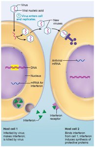
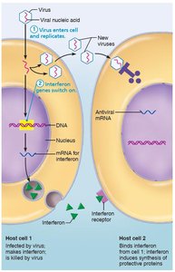
Interferons
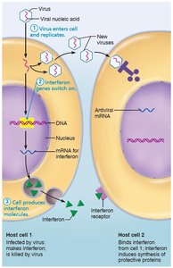
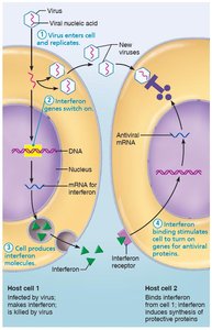
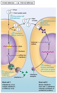
Interferons (IFNs) are proteins released by virus-infected cells that warn neighboring cells and stimulate production of antiviral proteins. Types include IFN-α, IFN-β, and IFN-γ, each with specific immune functions.
IFN-α and IFN-β: Block viral reproduction, activate NK cells.
IFN-γ: Activates macrophages, has widespread immune effects.
Clinical use: Artificial IFNs treat hepatitis C, genital warts, and multiple sclerosis.
Complement System
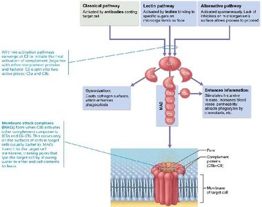
The complement system consists of about 20 blood proteins that circulate in inactive form. It can be activated by three pathways: classical, lectin, and alternative. All pathways converge on C3, leading to inflammation, opsonization, and cell lysis.
Classical pathway: Activated by antibodies binding to pathogens.
Lectin pathway: Activated by lectins binding to sugars on pathogens.
Alternative pathway: Activated spontaneously on pathogen surfaces.
Membrane attack complex (MAC): Forms holes in pathogen membranes, causing lysis.
C3b: Opsonizes pathogens, enhances phagocytosis.
C3a: Amplifies inflammation.
Fever
Fever is a systemic response to infection, initiated by pyrogens released by leukocytes and macrophages. It raises body temperature, inhibits microbial growth, and accelerates tissue repair.
Benefits: Sequesters iron and zinc, increases metabolic rate, enhances repair.
Summary Table: Innate Defenses
Category/Associated Elements | Protective Mechanism |
|---|---|
Phagocytes | Engulf and destroy pathogens; contribute to adaptive responses |
Natural killer (NK) cells | Induce apoptosis in abnormal cells; recognize general abnormality |
Inflammatory response | Prevents spread, disposes debris/pathogens, alerts adaptive immunity, promotes repair |
Antimicrobial proteins (Interferons, Complement) | Block viral reproduction, enhance phagocytosis, amplify inflammation |
Fever | High temperature inhibits microbes and enhances repair |
