Back
BackIntegrated Blueprint of the Muscular and Cardiovascular Systems: Structure, Function, and Physiology
Study Guide - Smart Notes
 Tailored notes based on your materials, expanded with key definitions, examples, and context.
The Biological Machine: Overview of Muscular and Cardiovascular Integration
This section introduces the concept of the human body as a biological machine, focusing on the integration of the muscular and cardiovascular systems. These systems work together to enable movement, maintain homeostasis, and support exercise physiology.
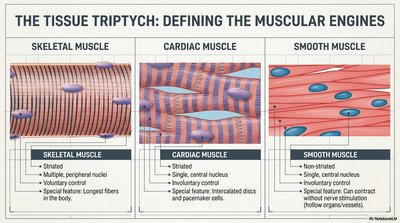
The Tissue Triptych: Defining the Muscular Engines
Skeletal Muscle
Skeletal muscle is responsible for voluntary movements and is attached to bones by tendons. It is characterized by its striated appearance and multiple peripheral nuclei.
Striated (striped appearance under microscope)
Voluntary control (conscious movement)
Multiple, peripheral nuclei
Longest fibers in the body
Cardiac Muscle
Cardiac muscle is found only in the heart and is responsible for pumping blood throughout the body. It is striated, has single central nuclei, and is under involuntary control.
Striated
Single, central nucleus
Involuntary control
Intercalated discs and pacemaker cells for synchronized contraction
Smooth Muscle
Smooth muscle is found in the walls of hollow organs and blood vessels. It is non-striated, has single central nuclei, and is under involuntary control.
Non-striated
Single, central nucleus
Involuntary control
Controls organ/vessel movement
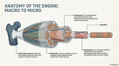
Anatomy of the Engine: Macro to Micro Structure of Skeletal Muscle
Skeletal muscle is organized from large to small structures, each with specific functions in force generation and movement.
Tendon/Aponeurosis: Connects muscle to bone, anchoring the muscle.
Epimysium: Dense outer layer separating muscle from other tissues.
Perimysium: Surrounds bundles (fascicles) of muscle fibers, containing blood vessels and nerves.
Endomysium: Surrounds individual muscle fibers, containing satellite cells for repair.
Myofibril: The contractile element within muscle fibers.
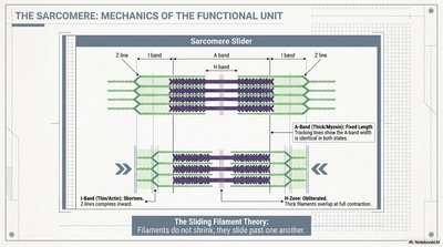
The Sarcomere: Mechanics of the Functional Unit
The sarcomere is the basic contractile unit of muscle fiber. Muscle contraction is explained by the sliding filament theory, where actin and myosin filaments slide past each other without changing length.
Z line: Defines the boundary of each sarcomere.
I band: Contains only thin (actin) filaments; shortens during contraction.
A band: Contains thick (myosin) filaments; remains constant in length.
H zone: Central region with only thick filaments; shortens during contraction.
Sliding Filament Theory: Filaments slide past each other to shorten the sarcomere and contract the muscle.
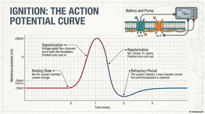
Ignition: The Action Potential Curve
Muscle contraction is initiated by an action potential, a rapid change in membrane potential that travels along the muscle fiber.
Resting State: Maintained by Na+-K+ pumps.
Depolarization: Voltage-gated Na+ channels open, Na+ rushes in.
Repolarization: Na+ channels close, K+ channels open, K+ exits.
Refractory Period: The cell cannot fire another impulse until baseline is restored.
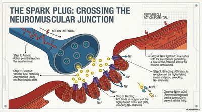
The Spark Plug: Crossing the Neuromuscular Junction
The neuromuscular junction is the synapse between a motor neuron and a muscle fiber, where the nerve impulse triggers muscle contraction.
Arrival: Action potential reaches the axon terminal.
Release: Vesicles release acetylcholine (ACh) into the synaptic cleft.
Binding: ACh binds to receptors on the muscle cell membrane, opening Na+ channels.
New Ignition: Na+ rushes in, generating a new action potential in the muscle.
Cleanup: Acetylcholinesterase (AChE) breaks down ACh, ending the signal.
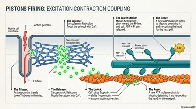
Pistons Firing: Excitation-Contraction Coupling
Excitation-contraction coupling describes the sequence from action potential arrival to muscle fiber contraction.
The Trigger: Action potential travels down T-tubules.
The Release: Sarcoplasmic reticulum releases Ca2+ into the cytosol.
The Unlock: Ca2+ binds to troponin, shifting tropomyosin and exposing actin sites.
The Power Stroke: Myosin heads bind to actin, pull, and release ADP + Pi.
The Reset: ATP binds to myosin, detaching it from actin and resetting the cycle.
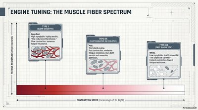
Engine Tuning: The Muscle Fiber Spectrum
Muscle fibers are classified based on contraction speed and fatigue resistance.
Type I (Slow Oxidative): High endurance, aerobic, fatigue-resistant, deep red color.
Type IIa (Fast Oxidative-Glycolytic): Intermediate endurance and power, uses both aerobic and anaerobic pathways.
Type IIx (Fast Glycolytic): Low endurance, high power, fatigues quickly, pale color.
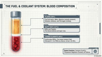
The Fuel & Coolant System: Blood Composition
Blood is a specialized connective tissue with multiple components, each serving distinct functions in transport, defense, and homeostasis.
Plasma (55%): Fluid matrix containing water, proteins (albumins, globulins, fibrinogen), and solutes.
Buffy Coat (1%): White blood cells and platelets for defense and repair.
Formed Elements (45%): Red blood cells (erythrocytes) for oxygen transport.
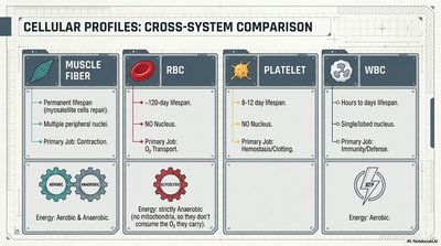
Cellular Profiles: Cross-System Comparison
Different cell types in muscle and blood have specialized structures and functions.
Cell Type | Lifespan | Nucleus | Primary Job | Energy |
|---|---|---|---|---|
Muscle Fiber | Permanent | Multiple, peripheral | Contraction | Aerobic & Anaerobic |
RBC | ~120 days | No | O2 Transport | Strictly Anaerobic |
Platelet | 8-12 days | No | Hemostasis/Clotting | --- |
WBC | Hours to days | Single/lobed | Immunity/Defense | Aerobic |
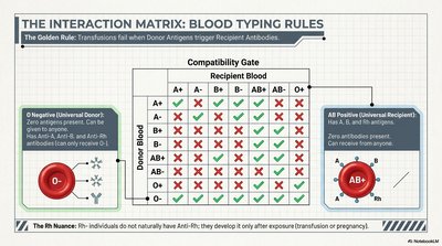
The Interaction Matrix: Blood Typing Rules
Blood transfusion compatibility depends on the presence or absence of antigens and antibodies. The golden rule is that transfusions fail when donor antigens trigger recipient antibodies.
O Negative: Universal donor (no A, B, or Rh antigens).
AB Positive: Universal recipient (no anti-A, anti-B, or anti-Rh antibodies).
Rh Nuance: Anti-Rh antibodies develop only after exposure.
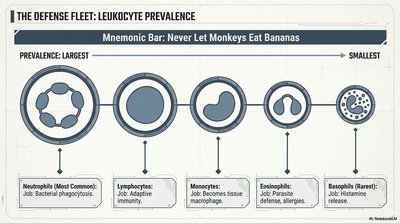
The Defense Fleet: Leukocyte Prevalence
Leukocytes (white blood cells) are classified by prevalence and function. The mnemonic "Never Let Monkeys Eat Bananas" helps recall the order: Neutrophils, Lymphocytes, Monocytes, Eosinophils, Basophils.
Neutrophils: Most common; bacterial phagocytosis.
Lymphocytes: Adaptive immunity.
Monocytes: Become tissue macrophages.
Eosinophils: Parasite defense, allergies.
Basophils: Rarest; histamine release.
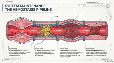
System Maintenance: The Hemostasis Pipeline
Hemostasis is the process of stopping bleeding and involves three main steps:
Vascular Spasm: Smooth muscle contracts to reduce blood flow.
Platelet Plug: Platelets adhere to exposed collagen and aggregate.
Coagulation: Fibrinogen is converted to fibrin, forming a stable clot.
Retraction & Fibrinolysis: Clot retracts and is eventually dissolved.
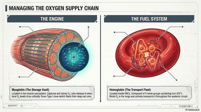
Managing the Oxygen Supply Chain
Oxygen is transported and stored by specialized proteins in muscle and blood.
Myoglobin: Stores O2 in muscle, releases it during exercise.
Hemoglobin: Transports O2 in blood, binds O2 in lungs and releases it in tissues.
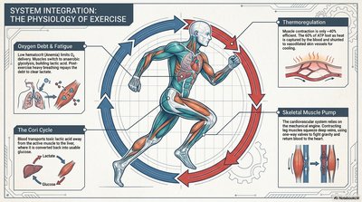
System Integration: The Physiology of Exercise
Exercise physiology involves the coordinated response of muscular and cardiovascular systems to meet increased metabolic demands.
Oxygen Debt & Fatigue: Low hemoglobin impairs O2 delivery, leading to fatigue.
The Cori Cycle: Lactate from muscle is converted back to glucose in the liver.
Thermoregulation: Muscle contraction generates heat; excess heat is dissipated by increased blood flow and sweating.
Skeletal Muscle Pump: Muscle contractions help return venous blood to the heart.