 Back
BackIntegrated CNS Function: Voluntary Motor Control
Study Guide - Smart Notes
 Tailored notes based on your materials, expanded with key definitions, examples, and context.
Integrated CNS Function: Voluntary Motor Control
Overview of Voluntary Motor Control
Voluntary motor control is the process by which the central nervous system (CNS) translates intentions and thoughts into coordinated skeletal muscle movements. This process involves multiple neural structures and pathways that work together to ensure smooth, purposeful, and adaptive actions.
Neural Components for Smooth Voluntary Movements
Steps in Voluntary Movement
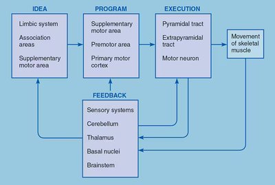
The successful execution of a voluntary motor task involves four main components:
Developing the idea to move: The intention or goal to move is formulated in higher brain centers, such as the prefrontal cortex, association areas, basal nuclei, and limbic system. This step is influenced by sensory input, memories, and emotions.
Programming motor commands: The motor plan is developed in the supplementary motor area, premotor cortex, and primary motor cortex. This involves selecting which muscles to activate and which to inhibit.
Executing the movement: Motor commands are sent via descending pathways to activate the appropriate motor neurons, resulting in muscle contraction.
Feedback and adjustment: Sensory feedback from the body and environment is integrated by the cerebellum, thalamus, basal nuclei, and brainstem to refine and adjust movements in real time.
Example: When reaching for a cup, the brain formulates the intention, programs the sequence of muscle activations, executes the movement, and uses sensory feedback to adjust the hand's trajectory.
Lateral Pathways: Control of Voluntary Movement
Pyramidal and Rubrospinal Tracts
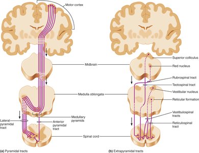
Lateral pathways are responsible for controlling fine, voluntary movements, especially of the distal extremities (hands and fingers). The two main tracts are:
Pyramidal tracts: Originate in the primary motor cortex and descend directly to the spinal cord. Most fibers cross to the opposite side at the medullary pyramids. They primarily control fine, discrete movements.
Rubrospinal tracts: Originate in the red nuclei of the midbrain, cross over, and join the pyramidal tract axons. They play a minor role in humans compared to the pyramidal tracts.
Example: Writing with a pen involves precise finger movements controlled by the pyramidal system.
Ventromedial Pathways: Control of Voluntary and Involuntary Movements
Vestibulospinal, Tectospinal, and Reticulospinal Tracts
Ventromedial pathways originate in the brainstem and are involved in the control of posture, balance, and gross movements of the trunk and proximal limbs. These pathways are indirect and do not synapse directly on motor neurons.
Vestibulospinal tracts: Originate in the vestibular nuclei and control muscles for posture and balance based on input from the inner ear.
Tectospinal tracts: Originate in the superior colliculi and control head and eye movements in response to visual and auditory stimuli.
Reticulospinal tracts: Originate in the reticular formation and regulate muscle tone and posture, with different branches stimulating or inhibiting extensor muscles.
Comparison: Lateral pathways (pyramidal and rubrospinal) control fine, distal movements, while ventromedial pathways control posture and gross movements.
The Control of Posture by the Brainstem
Brainstem Nuclei and Postural Adjustments
The brainstem contains nuclei that are part of the ventromedial pathways and are essential for involuntary control of posture. These nuclei integrate sensory information from proprioceptors, the vestibular system, eyes, and ears to maintain balance and adjust posture during movement or in response to external stimuli.
Example: Turning your head toward a sudden sound involves automatic postural adjustments mediated by the brainstem.
The Role of the Cerebellum in Motor Coordination
Cerebellar Function and Feedback
The cerebellum acts as a guidance system for movement, comparing intended actions with actual performance and making corrections as needed. It receives input from the cortex, basal nuclei, brainstem, spinal cord, and sensory systems, and sends feedback to the cortex via the thalamus.
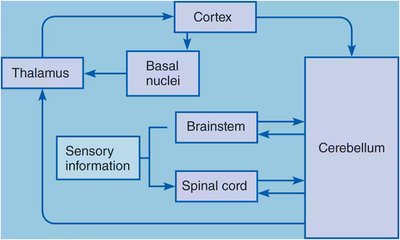
Maintains muscle tone and stores memories of motor activities for smoother execution of learned movements.
Damage to the cerebellum results in clumsy, uncoordinated movements and intention tremors.
Example: A person with cerebellar damage may have difficulty performing smooth, coordinated actions like reaching for a light switch.
The Basal Nuclei in Motor Control
Functions and Clinical Relevance
The basal nuclei (basal ganglia) provide feedback to the cortex for the development of motor strategies and the smoothing of movements. They are involved in the automatic execution of learned, repetitive motions and help inhibit unwanted movements.
Huntington's chorea: Characterized by loss of motor coordination and involuntary jerking movements due to overactive motor circuits.
Parkinson's disease: Involves loss of dopaminergic input to the basal nuclei, resulting in tremors, slow and stiff movements, and difficulty initiating movement.
Example: Difficulty in starting to walk or a shuffling gait in Parkinson's disease is due to basal nuclei dysfunction.
Summary Table: Functional Differences in Motor Pathways
Pathway | Origin | Main Function | Muscle Groups Controlled |
|---|---|---|---|
Lateral (Pyramidal, Rubrospinal) | Motor cortex, Red nucleus | Fine, voluntary movement | Distal extremities (hands, fingers) |
Ventromedial (Vestibulospinal, Tectospinal, Reticulospinal) | Brainstem nuclei | Posture, balance, gross movement | Trunk, neck, proximal limbs |
Key Structures in Motor Control: Roles
Structure | Role in Motor Control |
|---|---|
Cerebellum | Compares intended and actual movements; corrects errors; maintains muscle tone |
Basal Nuclei | Initiates and smooths movements; inhibits unwanted motions; involved in learned motor patterns |
Brainstem Nuclei | Controls posture and balance; integrates sensory input for postural adjustments |
Thalamus | Relays motor and sensory signals between cortex and other motor centers |
