 Back
BackIntegumentary and Skeletal Systems: Structure, Function, and Clinical Relevance
Study Guide - Smart Notes
 Tailored notes based on your materials, expanded with key definitions, examples, and context.
Chapter 5: The Integumentary System
Skin Structure and Layers
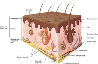
The skin, or integument, is the largest organ of the body and consists of two main layers: the superficial epidermis and the deeper dermis, which rests on the hypodermis (not part of the skin proper). The integument serves multiple functions, including protection, sensation, thermoregulation, excretion, and vitamin D synthesis.
Epidermis: Composed mainly of keratinocytes, with tactile cells (sensory receptors) and dendritic cells (immune function).
Dermis: Contains connective tissue, blood vessels, nerves, and accessory structures such as hair follicles and glands.
Hypodermis: Subcutaneous layer composed mainly of adipose tissue, providing insulation and cushioning.
Epidermal Layers
The epidermis is organized into distinct strata (layers), each with specialized cells and functions. From deep to superficial, the layers are:
Stratum basale: Single row of stem cells; contains keratinocytes, melanocytes (produce melanin), and tactile epithelial cells.
Stratum spinosum: Several layers of keratinocytes; contains dendritic cells for immune defense.
Stratum granulosum: 3–5 layers of flattened keratinocytes with cytoplasmic granules; cells begin to die.
Stratum lucidum: Thin, clear layer found only in thick skin (palms, soles); composed of dead keratinocytes.
Stratum corneum: Multiple layers of dead, flattened keratinocytes; provides a tough, protective barrier.
Thick skin contains all five strata and is found on the palms and soles. Thin skin lacks the stratum lucidum and has thinner layers overall.
Dermis Structure
The dermis is divided into two layers:
Papillary layer: Composed of loose areolar connective tissue; contains dermal papillae that interlock with epidermal ridges, increasing surface area for exchange of gases, nutrients, and waste.
Reticular layer: Dense irregular connective tissue; contains collagen fibers that form tension (cleavage) lines, important in surgical incisions.
Skin Pigmentation
Skin color is primarily determined by the pigment melanin, produced by melanocytes in the stratum basale. Melanin protects against UV radiation. Other pigments include:
Hemoglobin: Red pigment in blood, gives skin a pinkish hue.
Carotene: Yellow-orange pigment from diet, accumulates in the stratum corneum.
Changes in skin color can indicate pathology (e.g., cyanosis, jaundice, pallor).
Skin Glands and Associated Structures
Sweat (sudoriferous) glands: Four types—eccrine (most common, thermoregulation), apocrine (axillary/genital, odor), ceruminous (earwax), and mammary (milk).
Sebaceous glands: Produce sebum (oil) for lubrication and waterproofing; associated with hair follicles.
Burns and Clinical Considerations
First-degree burns: Affect only the epidermis; redness and minor pain.
Second-degree burns: Involve epidermis and part/all of dermis; blistering and pain.
Third-degree burns: Destroy epidermis, dermis, and deeper tissues; may not be painful initially due to nerve destruction; risk of dehydration and infection.
The rule of nines is used to estimate the percentage of body surface area affected by burns for fluid resuscitation:
Chapter 6: Bones and Bone Tissue
Bone Structure: Compact and Spongy Bone
Bones are composed of two main types of tissue:
Compact bone: Dense outer layer; provides strength and resistance to compression and twisting.
Spongy bone (cancellous bone): Inner honeycomb-like structure; contains trabeculae and houses bone marrow.
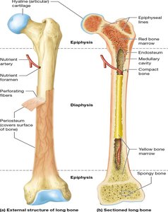
Long Bone Anatomy
Long bones have a characteristic structure:
Diaphysis: Shaft of the bone; surrounds the medullary (marrow) cavity.
Epiphyses: Rounded ends; covered with articular (hyaline) cartilage for joint movement.
Medullary cavity: Contains red or yellow bone marrow depending on age and bone type.
Periosteum: Dense irregular connective tissue membrane covering the bone; contains blood vessels and nerves.
Endosteum: Thin membrane lining the medullary cavity and trabeculae of spongy bone.
Sharpey's (perforating) fibers: Collagen fibers anchoring periosteum to bone.
Bone Matrix Composition
Inorganic matrix: Mainly calcium and phosphate salts (hydroxyapatite crystals), providing hardness and resistance to compression.
Organic matrix (osteoid): Predominantly collagen fibers, providing tensile strength and flexibility.
Hydroxyapatite formula:
Bone Cells
Osteoblasts: Bone-forming cells; synthesize osteoid.
Osteocytes: Mature bone cells; maintain bone matrix.
Osteoclasts: Bone-resorbing cells; break down bone matrix.
Bone Development and Growth
Ossification (osteogenesis): Process of bone formation, beginning in the embryonic period and continuing through childhood.
Intramembranous ossification: Bone develops from a membrane of embryonic connective tissue (e.g., flat bones of skull).
Endochondral ossification: Bone develops from a hyaline cartilage model (e.g., long bones).
Chapter 7: The Skeletal System
Overview of the Skeletal System
Axial skeleton: Skull, vertebral column, thoracic cage.
Appendicular skeleton: Pectoral girdle, upper limb, pelvic girdle, lower limb.
Bone markings: Depressions, openings, and projections for articulation and attachment.
The Skull
Cranial bones: Frontal, parietal (2), temporal (2), occipital, sphenoid, ethmoid.
Facial bones: Mandible, vomer, maxillary (2), zygomatic (2), nasal (2), lacrimal (2), palatine (2), inferior nasal conchae (2).
Sutures: Immovable joints connecting skull bones (except mandible).
Chapter 8: Articulations (Joints)
Classification of Joints
Functional classification:
Synarthrosis: Immovable (e.g., skull sutures).
Amphiarthrosis: Slightly movable (e.g., intervertebral discs).
Diarthrosis: Freely movable (e.g., synovial joints).
Structural classification:
Fibrous joints: Dense regular connective tissue; no joint cavity (e.g., sutures).
Cartilaginous joints: Cartilage connects bones; no joint cavity (e.g., synchondroses).
Synovial joints: Joint cavity filled with synovial fluid; most movable type (e.g., knee, hip).
Synovial Joint Structure and Movements
Articular cartilage: Hyaline cartilage covering bone ends.
Joint (synovial) cavity: Space containing synovial fluid.
Articular capsule: Encloses joint cavity; lined by synovial membrane.
Ligaments: Strengthen and support the joint.
Types of synovial joints: plane, hinge, pivot, condylar, saddle, ball-and-socket.
Common joint injuries include ligament tears (e.g., "unhappy triad" of the knee: tibial collateral ligament, medial meniscus, anterior cruciate ligament).
Examples of Joints
Atlantoaxial joint: Pivot joint between C1 and C2 vertebrae.
Glenohumeral joint: Ball-and-socket joint of the shoulder.
Intervertebral joint: Cartilaginous joint between vertebral bodies.
Interphalangeal joint: Hinge joint between phalanges.
Additional info: For a comprehensive understanding, students should refer to anatomical diagrams and models to visualize the structures described above. Clinical correlations, such as the effects of burns or joint injuries, are essential for applying anatomical knowledge to real-world scenarios.
