 Back
Integumentary System, Bone Structure, and Joints: ANP College Study Guide
Study Guide - Smart Notes
 Tailored notes based on your materials, expanded with key definitions, examples, and context.
The Integumentary System
Skin Layers and Structure
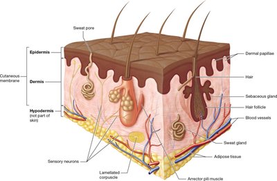
The skin is composed of three main layers: the superficial epidermis, the deeper dermis, and the underlying hypodermis. The integument serves several essential functions, including protection, sensation, thermoregulation, excretion, and vitamin D synthesis.
Epidermis: Contains mostly keratinocytes, which produce keratin. Also includes dendritic cells (immune function), melanocytes (produce melanin for UV protection), and tactile cells (sensory receptors).
Dermis: Consists of the papillary layer (areolar connective tissue, dermal papillae) and reticular layer (dense irregular connective tissue, tension lines).
Hypodermis: Not part of the skin proper; contains adipose tissue and anchors skin to underlying structures.
Epidermal Layers
The epidermis is organized into distinct strata, from deep to superficial:
Stratum basale: Single row of stem cells; contains keratinocytes, melanocytes, and tactile cells.
Stratum spinosum: Several layers of keratinocytes; contains dendritic cells.
Stratum granulosum: 3-5 layers of flattened keratinocytes with granules.
Stratum lucidum: Only in thick skin; thin layer of dead keratinocytes.
Stratum corneum: Many layers of dead, flattened keratinocytes.
Thick skin has all five strata and is found on palms and soles. Thin skin lacks the stratum lucidum.
Skin Pigmentation
Skin color is primarily determined by the pigment melanin, produced by melanocytes in the stratum basale. Other pigments include hemoglobin and carotene. Melanin protects against UV radiation and is synthesized from the amino acid tyrosine via the enzyme tyrosinase in melanosomes.
Melanin: Ranges from orange-red to black; protects DNA from UV damage.
Hemoglobin: Red pigment in blood; gives skin a pinkish hue.
Carotene: Yellow-orange pigment from diet; accumulates in stratum corneum.
Skin Glands
The skin contains two main types of glands:
Sweat (Sudoriferous) Glands: Four types—eccrine (most common, thermoregulation), apocrine (associated with hair follicles, odor), ceruminous (ear wax), and mammary (milk production).
Sebaceous Glands: Produce sebum (oil) for lubrication and protection; associated with hair follicles.
Burns and Skin Pathology
Burns are classified by depth:
First-degree: Only epidermis; minor pain and erythema.
Second-degree: Epidermis and part/all of dermis; pain, blistering, possible scarring.
Third-degree: Epidermis, dermis, hypodermis, and deeper tissues; major tissue damage, risk of dehydration and infection.
The rule of nines is used to estimate burn surface area for fluid replacement: each major body region represents 9% of total surface area (plus 1% for genital area).
Bone Structure and Classification
Long Bone Anatomy
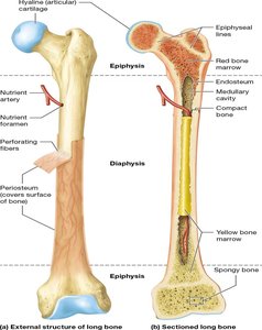
Long bones have a shaft (diaphysis) and two ends (epiphyses). The diaphysis surrounds the medullary cavity, which contains bone marrow. The epiphyses are covered with articular cartilage for joint movement.
Periosteum: Dense irregular connective tissue membrane covering bone; contains blood vessels and nerves.
Compact bone: Dense outer layer; resists compression and twisting.
Spongy bone: Inner honeycomb structure; contains trabeculae and bone marrow.
Bone Matrix and Cells
The bone matrix consists of:
Inorganic matrix: Mainly hydroxyapatite crystals ; provides strength and resistance to compression.
Organic matrix (osteoid): Collagen fibers, proteoglycans, glycoproteins; provides flexibility and resistance to tension.
Bone cells include:
Osteoblasts: Build new bone matrix.
STEP 1: Osteoblasts develop in the primary ossification center from mesenchymal cells. At the primary ossification center, mesenchymal cells differentiate first into osteogenic cells and then into osteoblasts.
STEP 2: Osteoblasts secrete organic matrix, which calcifies, and trapped osteoblasts become osteocytes. The newly formed osteoblasts secrete the organic matrix of bone. In a few days’ time, calcium salts and other components of the inorganic matrix are deposited in the primary ossification center, a process called calcification, and the early bone hardens. Trapped osteoblasts then become osteocytes.
STEP 3: Osteoblasts lay down trabeculae of early spongy bone, and some of the surrounding mesenchyme differentiates into the periosteum. Osteoblasts continue to lay down new bone, forming the trabeculae of the early spongy bone. Over time, the trabeculae enlarge and merge, forming larger trabeculae. About this time, some of the mesenchyme surrounding the developing bone differentiates into the periosteum. Some of the vascular tissue in the forming spongy bone will become bone marrow.
STEP 4: Osteoblasts in the periosteum lay down early compact bone. Osteoblasts within the periosteum continue to secrete organic bone matrix. This matrix becomes more heavily calcified than the dee
Osteocytes: Mature bone cells in lacunae; maintain bone tissue.
Osteoclasts: Resorb bone matrix.
Feature | Red Bone Marrow | Yellow Bone Marrow |
|---|---|---|
Primary Function | Blood cell production (hematopoiesis). | Energy storage (triglycerides). |
Main Components | Reticular fibers and blood-forming cells. | Blood vessels and adipocytes (fat cells). |
Prevalence in Children | Dominant type due to rapid growth. | Starts replacing red marrow around age 5. |
Prevalence in Adults | Limited; found only in select bones. | Dominant type throughout most of the body. |
Adult Locations | Pelvis, proximal femur/humerus, vertebrae, ribs, sternum, clavicles, scapulae, skull. | Most bones of the adult skeleton. |
Adaptability | Can be transplanted to treat blood diseases. | Can convert back to red marrow if blood cell production needs to increase. |
Bone Classification by Shape
Long bones: Longer than wide (e.g., femur).
Short bones: About as long as wide (e.g., carpals).
Flat bones: Broad, flat, thin (e.g., sternum).
Irregular bones: Complex shapes (e.g., vertebrae).
Sesamoid bones: Round, flat, within tendons (e.g., patella).
Bone Formation (Ossification)
Bone formation occurs via two pathways:
Intramembranous ossification: Forms flat bones from mesenchymal membrane.
Endochondral ossification: Forms most bones from hyaline cartilage model.
Primary bone (woven) is immature and weak; secondary bone (lamellar) is mature and strong.
Joints and Articulations
Joint Classification and Structure
Joints (articulations) enable movement, provide stability, and allow bone growth. They are classified functionally and structurally:
Functional: Synarthrosis (no movement), amphiarthrosis (some movement), diarthrosis (freely movable).
Structural: Fibrous (dense connective tissue), cartilaginous (cartilage), synovial (fluid-filled cavity).
Synovial Joints
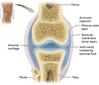
Synovial joints are characterized by a joint cavity filled with synovial fluid, articular cartilage, and a capsule with fibrous and synovial layers. They allow a wide range of movements and are the most common joint type in the body.
Synovial fluid: Lubricates, nourishes, and absorbs shock.
Articular cartilage: Reduces friction and absorbs impact.
Joint capsule: Provides structural support and encloses the joint cavity.
Types of Synovial Joints
Plane (gliding) joints: Flat surfaces, allow sliding movements (e.g., intercarpal joints).
Hinge joints: Uniaxial, allow flexion and extension (e.g., elbow).
Pivot joints: Uniaxial, allow rotation (e.g., atlantoaxial joint).
Other types: Condyloid, saddle, ball-and-socket joints allow various movements.
Fibrous and Cartilaginous Joints
Sutures: Immovable joints between skull bones.
Gomphoses: Joints between teeth and alveolar sockets.
Syndesmoses: Bones joined by interosseous membrane (e.g., radius and ulna).
Synchondroses: Bones held by hyaline cartilage (e.g., costochondral joints).
Symphyses: Bones joined by fibrocartilage (e.g., intervertebral discs).
Joint Pathology and Injury
Common joint injuries include ligament tears (e.g., "Unhappy Triad" in the knee), meniscus damage, and dislocations. Treatment often involves surgery and rehabilitation.
Table: Comparison of Joint Types
Type | Structural Feature | Movement | Example |
|---|---|---|---|
Fibrous | Dense connective tissue | Immovable or slightly movable | Sutures, syndesmoses |
Cartilaginous | Cartilage | Immovable or slightly movable | Synchondroses, symphyses |
Synovial | Joint cavity, synovial fluid | Freely movable | Knee, shoulder |
Additional info: Academic context was added to clarify bone matrix composition, ossification processes, and joint classification for completeness.
