 Back
Integumentary System: Pre-Lecture and Concept Check Guidance
Study Guide - Smart Notes
 Tailored notes based on your materials, expanded with key definitions, examples, and context.
Q1. What are the body’s most abundant epithelial cells?
Background
Topic: Integumentary System – Epidermal Cell Types
This question tests your understanding of the main cell types found in the epidermis, the outermost layer of the skin.
Key Terms:
Keratinocytes: The primary cell type in the epidermis, responsible for producing keratin, a protein that provides strength and waterproofing.
Melanocytes: Cells that produce the pigment melanin.
Desmosomes: Structures that connect cells together (not a cell type).
Gap junctions: Channels between cells (not a cell type).
Step-by-Step Guidance
Review the main cell types found in the epidermis and their functions.
Identify which cell type is most numerous and what its primary role is in the skin.
Eliminate options that are not cell types (e.g., desmosomes, gap junctions).
Try solving on your own before revealing the answer!
Q2. What are the cells that produce pigment?
Background
Topic: Integumentary System – Pigment Production
This question focuses on the cells responsible for producing melanin, the pigment that gives skin its color and protects against UV radiation.
Key Terms:
Melanocytes: Specialized cells in the epidermis that synthesize melanin.
Keratinocytes: Main structural cells, do not produce pigment.
Step-by-Step Guidance
Recall which cell type is responsible for pigment production in the skin.
Consider the function of each option and match it to pigment synthesis.
Eliminate options that do not produce pigment.
Try solving on your own before revealing the answer!
Q3. Which vitamin is converted from a steroid when epidermal cells are exposed to UV radiation and helps in the absorption of calcium and phosphorus?
Background
Topic: Integumentary System – Vitamin Synthesis
This question tests your knowledge of how the skin contributes to vitamin synthesis, specifically the vitamin involved in calcium and phosphorus absorption.
Key Terms:
Vitamin D: Produced in the skin from a precursor steroid when exposed to UV light; essential for calcium and phosphorus absorption.
Step-by-Step Guidance
Recall which vitamin is synthesized in the skin upon UV exposure.
Identify the vitamin’s role in mineral absorption.
Eliminate vitamins that are not involved in this process.
Try solving on your own before revealing the answer!
Q4. The dermis has two major layers. Which is the DEEPER of the two?
Background
Topic: Integumentary System – Dermal Structure
This question asks you to identify the deeper layer of the dermis, which lies beneath the epidermis and supports skin structure.
Key Terms:
Papillary layer: The superficial, upper layer of the dermis.
Reticular layer: The deeper, thicker layer of the dermis.
Step-by-Step Guidance
Recall the two main layers of the dermis and their relative positions.
Identify which layer is closer to the hypodermis (subcutaneous tissue).
Eliminate options that are not actual dermal layers.
Try solving on your own before revealing the answer!
Q5. Which of the following is one of the functions of the integumentary system?
Background
Topic: Integumentary System – Functions
This question tests your understanding of the main roles of the skin, such as protection, sensation, and water balance.
Key Terms:
Reduction of water loss: The skin acts as a barrier to prevent dehydration.
Step-by-Step Guidance
Review the primary functions of the integumentary system.
Identify which option matches a known function of the skin.
Eliminate options that are not functions of the integumentary system.
Try solving on your own before revealing the answer!
Q6. In which layer of the epidermis do cells stop dividing and start making large amounts of keratin?
Background
Topic: Integumentary System – Epidermal Layers
This question focuses on the process of keratinization and the specific layer where cells transition from dividing to producing keratin.
Key Terms:
Stratum granulosum: The layer where keratin production begins and cell division ceases.
Step-by-Step Guidance
Recall the order of epidermal layers from deep to superficial.
Identify the layer where cells stop dividing and begin keratinization.
Eliminate layers where cell division still occurs or where cells are already dead.
Try solving on your own before revealing the answer!
Q7. Subcutaneous injections are made in the:
Background
Topic: Integumentary System – Skin Layers and Clinical Application
This question tests your knowledge of the anatomical location used for subcutaneous injections, which is important for drug delivery.
Key Terms:
Hypodermis (subcutaneous layer): The layer beneath the dermis, rich in fat and blood vessels, used for subcutaneous injections.
Step-by-Step Guidance
Review the structure of the skin and the location of each layer.
Identify which layer is targeted for subcutaneous injections.
Eliminate layers that are not used for this purpose.
Try solving on your own before revealing the answer!
Q8. Which of the following is NOT an appendage of skin?
Background
Topic: Integumentary System – Skin Appendages
This question asks you to distinguish between true skin appendages and other body structures.
Key Terms:
Skin appendages: Structures derived from the epidermis, such as hair, nails, and glands.
Step-by-Step Guidance
List the main appendages of the skin.
Identify which option does not fit the definition of a skin appendage.
Eliminate options that are true appendages.
Try solving on your own before revealing the answer!
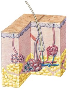
Q9. Apocrine sweat glands: Structure and Function
Background
Topic: Integumentary System – Sweat Glands
This section describes the characteristics of apocrine sweat glands, including their location, secretion type, and role in body odor.
Key Terms:
Apocrine glands: Large sweat glands that open into hair follicles in specific regions and produce a lipid-rich secretion.
Step-by-Step Guidance
Review the distinguishing features of apocrine glands (location, secretion, function).
Compare apocrine glands to other sweat glands (e.g., merocrine/eccrine glands).
Understand how apocrine gland activity contributes to body odor.
Try solving on your own before revealing the answer!
Q10. Contraction of the arrector pili muscles results in secretion from which gland type?
Background
Topic: Integumentary System – Glandular Secretion and Muscle Action
This question tests your understanding of the relationship between arrector pili muscle contraction and glandular secretion in the skin.
Key Terms:
Arrector pili muscle: Small muscles attached to hair follicles that cause "goosebumps" when contracted.
Sebaceous glands: Oil glands associated with hair follicles, often stimulated by arrector pili contraction.
Step-by-Step Guidance
Recall the function of the arrector pili muscle and its association with hair follicles.
Identify which gland type is stimulated by this muscle’s contraction.
Eliminate gland types not associated with hair follicles or arrector pili muscles.
Try solving on your own before revealing the answer!
