Back
BackIntegumentary System: Skin, Hair, Glands, and Nails
Study Guide - Smart Notes
 Tailored notes based on your materials, expanded with key definitions, examples, and context.
Skin Color Variation
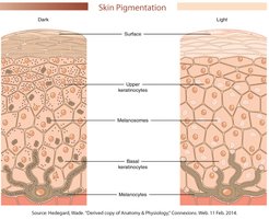
Melanin and Skin Pigmentation
Skin color is primarily determined by the type and distribution of melanin, a pigment produced by melanocytes in the epidermis. There are two main types of melanin: eumelanin (brown-black pigment) and pheomelanin (yellow-red pigment). The amount and location of melanin in the skin layers contribute to the visible differences in skin color among individuals.
Lighter skin: Contains more pheomelanin, which tends to remain in the basal layers and is lost in higher layers of the epidermis.
Darker skin: Contains more eumelanin, which is produced in greater quantities and persists throughout all layers of the epidermis.
Skin Color as a Diagnostic Tool
Clinical Significance of Skin Color Changes
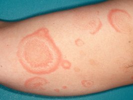
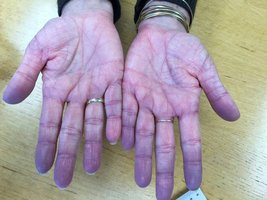
Changes in skin color can indicate underlying physiological or pathological conditions, often related to blood flow or oxygenation in the dermis.
Erythema: Reddening of the skin due to increased dermal blood flow, commonly seen with exercise, trauma, fever, or infection.
Pallor: Paleness of the skin resulting from decreased blood flow, often observed in cold conditions or during the 'fight or flight' response.
Cyanosis: Bluish discoloration of the skin caused by low oxygen levels in the blood, which may occur with respiratory difficulties or low hemoglobin levels.
Vitamin D Synthesis and Skin
Role of Sunlight in Vitamin D Production
Exposure to sunlight enables epidermal cells to convert a steroid precursor into vitamin D3 (cholecalciferol). This process is essential for calcium homeostasis and bone health.
UV light stimulates the synthesis of vitamin D3 in the skin.
Vitamin D3 is then converted by the liver and kidneys into calcitriol, the active form of vitamin D.
Calcitriol promotes the absorption of calcium ions (Ca2+) in the small intestine, which is critical for bone mineralization.
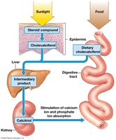
Clinical Note: Insufficient sunlight exposure or dietary intake of vitamin D can lead to bone disorders such as rickets or osteomalacia.
Hair Structure and Function
Hair Anatomy and Growth
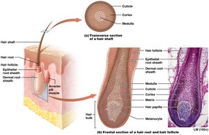
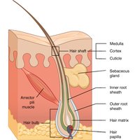
Hair is a nonliving accessory structure of the skin, composed of keratinized cells produced in hair follicles. It serves protective, insulating, and sensory functions.
Shaft: The visible part of the hair, consisting of dead, keratinized cells.
Root: The portion below the skin surface, beginning at the hair bulb and surrounded by the root sheath.
Hair papilla: Contains blood vessels and nerves at the base of the follicle, supporting hair growth.
Medulla, cortex, cuticle: The medulla contains soft keratin, the cortex contains hard keratin, and the cuticle is the outermost layer of dead cells.
Goosebumps and Arrector Pili Muscles
Arrector pili muscles are small muscles attached to hair follicles. Their contraction causes hair to stand up (piloerection), producing 'goosebumps' in response to cold or emotional stimuli.
Types and Texture of Hair
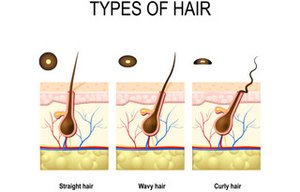
Hair texture and type are determined by the shape of the hair follicle and the amount of keratin produced.
Lanugo: Fine, non-pigmented hair covering the fetus, replaced before birth.
Vellus hair: Thin, non-pigmented 'peach fuzz' found on children and adults.
Terminal hair: Thick, coarse, pigmented hair found on the scalp, eyebrows, and after puberty in other regions.
Hair texture: Straight hair has round follicles, curly hair has oval follicles.
Hair Pigmentation
Hair color is determined by the type and amount of melanin produced by melanocytes in the hair follicle.
Blond hair: Contains little melanin.
Black hair: Contains abundant eumelanin.
Red hair: Contains pheomelanin.
Gray/white hair: Results from decreased melanin production with age.
Glands of the Skin
Types of Skin Glands
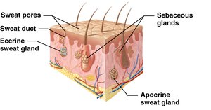
The skin contains two main types of exocrine glands: sweat (sudoriferous) glands and sebaceous glands. Both are derived from epidermal epithelial cells but are located in the dermis.
Sweat glands: Produce sweat for thermoregulation and waste excretion.
Sebaceous glands: Secrete oily sebum to lubricate and protect the skin and hair.
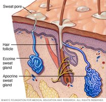
Sweat (Sudoriferous) Glands
There are four types of sweat glands, all using merocrine secretion (exocytosis):
Eccrine sweat glands: Most common; secrete watery sweat directly onto the skin surface for cooling (sensible perspiration).
Apocrine sweat glands: Found in axillae and pubic regions; secrete a thicker, cloudy fluid into hair follicles, which becomes odorous when metabolized by skin bacteria. Active after puberty.
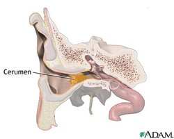
Ceruminous glands: Modified sweat glands in the ear canal that produce cerumen (ear wax) to trap particles.
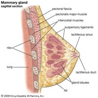
Mammary glands: Highly specialized glands that produce milk in mammals.
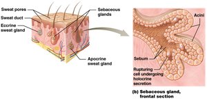
Sebaceous (Oil) Glands
Sebaceous glands secrete sebum, a waxy or oily lipid, into hair follicles or directly onto the skin surface. Sebum lubricates and protects the skin and hair, and inhibits the growth of certain bacteria. These glands are most abundant on the face and scalp and are influenced by sex hormones, especially during puberty.
Holocrine secretion: Sebum is released by the rupture of gland cells.
Distribution: Absent on palms and soles.
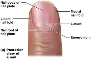
Nails
Structure and Function of Nails
Nails are hard, keratinized structures that protect the tips of fingers and toes. They are composed of stratified squamous epithelium filled with keratin.
Nail plate: The main body of the nail, sitting atop the nail bed.
Nail body: The visible portion of the nail plate.
Nail root: The portion under the skin where actively dividing cells are found.
Cuticle (eponychium): The fold of skin covering the nail root.
Skin Response to Injury
Wound Healing and Repair
The skin responds to injury through a series of steps involving inflammation, cell migration, proliferation, and remodeling to restore tissue integrity. The process includes clot formation, re-epithelialization, and scar formation if the damage is extensive.