 Back
BackIntegumentary System: Structure, Function, and Clinical Relevance
Study Guide - Smart Notes
 Tailored notes based on your materials, expanded with key definitions, examples, and context.
Integumentary System
Overview and Functions
The integumentary system consists of the skin and its accessory organs, including hair, nails, and cutaneous glands. It serves as the body's primary barrier to the external environment and plays vital roles in protection, sensation, thermoregulation, and more.
Protection: Acts as a physical, chemical, and physiological barrier against trauma, infection, and dehydration. Keratin, acidic secretions, and immune cells (Langerhans cells) contribute to defense.
Excretion: Removes nitrogenous waste through sweat.
Vitamin D Synthesis: Initiates vitamin D production, completed by the liver and kidneys.
Sensation: Contains receptors for temperature, touch, and pain.
Thermoregulation: Maintains body temperature via vasoconstriction, vasodilation, and perspiration.
Nonverbal Communication: Facial expressions and skin appearance contribute to social interactions.
Structure of the Skin
Layers of the Skin
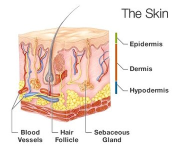
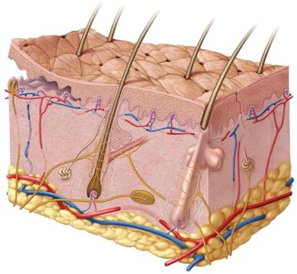
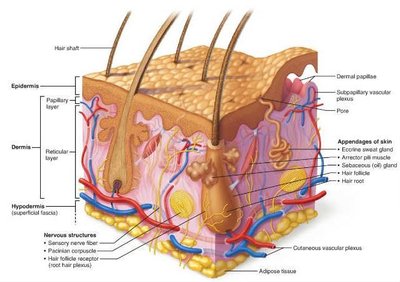
The skin is the largest organ of the body, comprising three main layers:
Epidermis: Outermost layer; keratinized stratified squamous epithelium. Lacks blood vessels and relies on diffusion from the dermis.
Dermis: Middle layer; composed of connective tissue, provides strength and elasticity.
Hypodermis (Subcutaneous Layer): Not technically part of the skin, but closely associated. Contains areolar and adipose tissue, serving as energy storage and insulation.
Epidermis: Cell Types and Layers
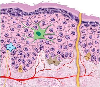
The epidermis contains several specialized cell types and is organized into distinct layers:
Stem Cells: Undifferentiated cells in the stratum basale that give rise to keratinocytes.
Keratinocytes: Most abundant; produce keratin for protection.
Melanocytes: Produce melanin pigment, protecting DNA from UV radiation.
Merkel (Tactile) Cells: Sensory receptors for touch.
Langerhans (Dendritic) Cells: Immune cells that guard against pathogens.
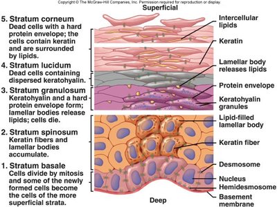
The main layers (from deep to superficial):
Stratum basale: Basal layer with stem cells, melanocytes, and Merkel cells.
Stratum spinosum, granulosum, lucidum (in thick skin), and stratum corneum: Surface layer of dead, keratinized cells providing barrier function.
Keratinization and Life Cycle of Keratinocytes
Keratinocytes originate in the stratum basale and migrate upward, accumulating keratin and eventually dying. The process from production to exfoliation takes 30–40 days, faster in injured or stressed skin.
Calluses/Corns: Thickened accumulations of dead keratinocytes due to friction.
Dermis: Structure and Zones
The dermis is a connective tissue layer beneath the epidermis, providing structural support and housing blood vessels, nerves, and accessory structures.
Papillary Layer: Superficial, areolar tissue with dermal papillae that interlock with epidermal ridges, forming fingerprints.
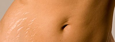
Reticular Layer: Deeper, dense irregular connective tissue; contains collagen fibers. Stretch marks (striae) result from tearing of these fibers.
Hypodermis (Subcutaneous Tissue)
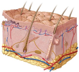
The hypodermis contains more areolar and adipose tissue than the dermis, serving as an energy reservoir and providing thermal insulation. It is a common site for drug injection due to its vascularity.
Skin Color and Markings
Determinants of Skin Color
Skin color is primarily determined by melanin, produced by melanocytes and distributed to keratinocytes. Other pigments and factors also contribute:
Melanin: Exists as eumelanin (brown-black) and pheomelanin (red-yellow). Darker skin results from more melanin production and slower breakdown.
Hemoglobin: Red pigment in blood, gives skin a pinkish hue.
Carotene: Yellow pigment from diet, accumulates in stratum corneum and fat.

Clinical and Diagnostic Skin Colors
Cyanosis: Blue tint due to low oxygen.
Erythema: Redness from increased blood flow.
Pallor: Paleness from reduced blood flow.
Albinism: Genetic lack of melanin.
Jaundice: Yellowing from bilirubin accumulation.
Hematoma: Bruising from clotted blood under skin.
Skin Markings
Freckles: Flat, melanized patches.
Moles (Nevi): Elevated, melanized patches, sometimes with hair.
Hemangiomas: Benign tumors of dermal capillaries (birthmarks).
Accessory Structures: Hair and Nails
Hair (Pilus): Structure and Function
Hair is a filament of keratinized cells growing from a follicle. It is found almost everywhere except palms, soles, lips, and certain genital areas.
Zones: Bulb (base, living cells), root (within follicle), shaft (above skin).
Layers: Medulla (core), cortex (bulk), cuticle (outer layer).
Follicle: Includes epithelial and connective tissue sheaths, hair receptors, and piloerector muscle (causes goosebumps).
Hair Texture and Color
Texture: Determined by cross-sectional shape (round = straight, oval = wavy, flat = curly).
Color: Eumelanin (brown/black), pheomelanin (red/yellow), little or no melanin (gray/white).
Functions of Hair
Vestigial in humans, but provides sensory input, heat retention, and protection (e.g., eyelashes, guard hairs in nostrils).
Pubic and axillary hair signal sexual maturity and aid in scent dispersal.
Nails: Structure and Function
Nail Plate: Hard part, includes free edge, nail body, and nail root.
Functions: Aid in manipulation, protect fingertips, and enhance sensation.
Skin Glands
Types of Exocrine Glands
Sebaceous Glands: Secrete sebum (oily), lubricates hair and skin.
Sudoriferous (Sweat) Glands: Regulate temperature and excrete waste.
Ceruminous Glands: Produce cerumen (earwax) for ear canal protection.
Mammary Glands: Secrete milk.
Clinical Correlations: Skin Disorders
Skin Cancer
Basal Cell Carcinoma: Most common, least dangerous, arises from stratum basale.
Squamous Cell Carcinoma: Arises from keratinocytes of stratum spinosum, can metastasize.
Malignant Melanoma: Most deadly, arises from melanocytes, high risk with family history and severe childhood sunburns.
Burns
First-degree: Only epidermis, redness and pain, heals quickly.
Second-degree: Epidermis and part of dermis, blisters, may scar.
Third-degree: Full thickness, destroys epidermis and dermis, requires grafts, risk of fluid loss and infection.
Summary Table: Layers of the Skin
Layer | Main Tissue | Key Features |
|---|---|---|
Epidermis | Keratinized stratified squamous epithelium | No blood vessels, multiple strata, protective barrier |
Dermis | Connective tissue (areolar and dense irregular) | Blood vessels, nerves, glands, hair follicles |
Hypodermis | Areolar and adipose tissue | Energy storage, insulation, vascular |
