 Back
BackIntegumentary System: Structure, Function, and Clinical Relevance
Study Guide - Smart Notes
 Tailored notes based on your materials, expanded with key definitions, examples, and context.
Functional Anatomy of the Skin
Overview of the Integumentary System
The integumentary system is the body's most accessible organ system, comprising the skin and its accessory structures. It serves as the first line of defense against environmental threats and plays a crucial role in homeostasis.
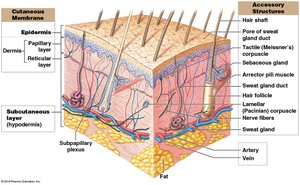
Cutaneous membrane: Includes the epidermis (stratified squamous epithelium) and dermis (papillary and reticular layers).
Accessory structures: Hair, nails, exocrine glands, sensory receptors, arrector pili muscles, and cutaneous plexus.
Subcutaneous layer (hypodermis): Not part of the skin, but separates it from deeper tissues and stores fat.
Functions of the Integumentary System
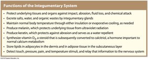
The integumentary system performs several essential functions for the body.
Protection: Shields underlying tissues from impact, abrasion, fluid loss, and chemical attack.
Excretion: Removes salts, water, and organic wastes via glands.
Temperature regulation: Maintains normal body temperature through insulation or evaporative cooling.
Keratin production: Provides abrasion resistance and water repellency.
Vitamin D3 synthesis: Enables calcium metabolism.
Lipid storage: Stores lipids in dermis and subcutaneous layer.
Sensory detection: Detects touch, pressure, pain, and temperature.
Structural Features of the Epidermis
Epidermal Layers and Their Functions
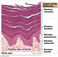
The epidermis is composed of multiple layers (strata) of keratinocytes, each with distinct functions.
Stratum basale: Deepest layer, attached to basement membrane; contains basal cells (stem cells) and Merkel cells (touch receptors).
Stratum spinosum: 8–10 layers of keratinocytes; contains dendritic (Langerhans) cells for immune defense.
Stratum granulosum: 3–5 layers; cells produce keratin and keratohyalin, become thinner and less permeable.
Stratum lucidum: Only in thick skin; densely packed dead cells filled with keratin.
Stratum corneum: Outermost layer; 15–30 layers of dead, keratinized cells; water resistant.
Thin Skin vs. Thick Skin
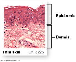
Thin skin: Covers most of the body; contains four strata.
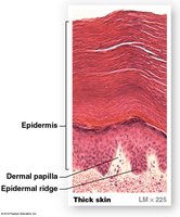
Thick skin: Found on palms and soles; contains five strata, including stratum lucidum.
Epidermal Ridges and Fingerprints
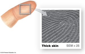
Epidermal ridges: Deep layers of epidermis form ridges adjacent to dermal papillae, increasing surface area for attachment.
Fingerprints: Unique patterns of epidermal ridges used for identification.
Skin Color and Clinical Relevance
Factors Influencing Skin Color
Skin color is determined by epidermal pigmentation and dermal circulation.
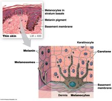
Carotene: Orange-yellow pigment.
Melanin: Brown/black or red/yellow pigment produced by melanocytes; packaged into melanosomes and transferred to keratinocytes.
Hemoglobin: Red pigment in blood; increased blood flow causes redness, decreased flow causes pallor or cyanosis.
UV exposure: Increases melanin production.
Skin Cancer Types
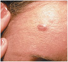
Basal cell carcinoma: Most common; originates in stratum basale; rarely metastasizes.
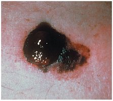
Malignant melanoma: Dangerous; cancerous melanocytes metastasize rapidly; early detection is critical.
Dermis and Subcutaneous Layer
Dermis Structure and Function
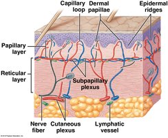
The dermis lies between the epidermis and hypodermis, providing structural support and housing blood vessels, nerves, and accessory organs.
Papillary layer: Areolar tissue; contains capillaries, lymphatic vessels, and sensory neurons.
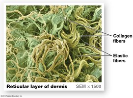
Reticular layer: Dense irregular connective tissue; contains collagen and elastic fibers, blood vessels, and accessory organs.
Subcutaneous Layer (Hypodermis)
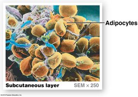
Adipose tissue: Dominates hypodermis; important for energy storage and insulation.
Fat distribution: Varies by sex and age; more in abdominal region, less in hands and feet.
Sensory Receptors in Skin
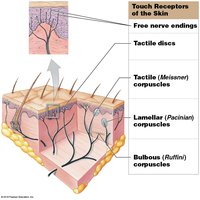
Free nerve endings: Sensitive to touch and pressure.
Tactile (Merkel) discs: Detect texture and light pressure.
Meissner corpuscles: Detect light touch, pressure, and vibration.
Pacinian corpuscles: Detect deep pressure and vibration.
Ruffini corpuscles: Sensitive to pressure and stretching.
Tension (Cleavage) Lines
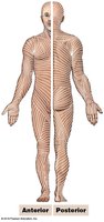
Formed by collagen and elastic fiber arrangement: Important for surgical incisions and wound healing.
Parallel cuts: Heal better, less scarring.
Perpendicular cuts: More scarring.
Clinical Module: Burns and Skin Grafts
Classification of Burns
Burns are classified by depth and area affected.
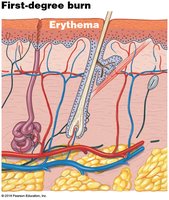
First-degree: Only epidermis; redness and inflammation.
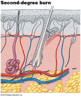
Second-degree: Epidermis and part of dermis; blistering, pain, swelling.
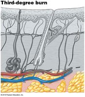
Third-degree: Epidermis, dermis, and subcutaneous layer; less pain, requires skin grafting.
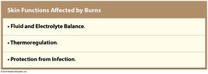
Skin Functions Affected by Burns
Fluid and electrolyte balance
Thermoregulation
Protection from infection
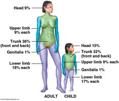
Rule of Nines
Used to estimate percentage of body surface area affected by burns.
Modified for children due to different body proportions.
Types of Skin Grafts
Split-thickness: Epidermis and superficial dermis.
Full-thickness: Epidermis and both dermal layers.
Autograft: Patient's own skin; best choice, no rejection.
Allograft: Cadaver skin.
Xenograft: Animal skin.
Accessory Structures of the Skin
Hair, Nails, and Exocrine Glands
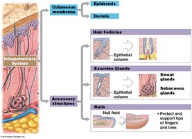
Accessory structures are epidermal derivatives that originate from the epidermis during development.
Hair follicles: Produce hair for protection and sensation.
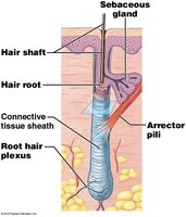
Exocrine glands: Sweat glands (thermoregulation, waste excretion), sebaceous glands (lubrication).
Nails: Protect and support finger and toe tips.
Mechanisms of Hair Production
Hair Structure and Growth
Hair is composed of dead, keratinized cells produced in specialized follicles.
Terminal hairs: Large, coarse, pigmented (scalp, armpit).
Vellus hairs: Small, short, delicate (general body surface).
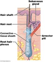
Hair shaft: Visible part above skin.
Hair root: Anchors hair in skin.
Root hair plexus: Sensory nerves at follicle base.
Arrector pili: Smooth muscle causing hair to stand erect.
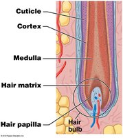
Hair bulb: Expanded base surrounding hair papilla (blood vessels, nerves).
Hair matrix: Basal cells actively dividing.
Hair Structure
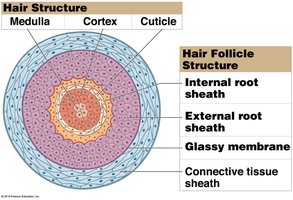
Medulla: Core with soft keratin.
Cortex: Thick layer with hard keratin.
Cuticle: Surface layer with hard keratin.
Internal root sheath: Surrounds root and shaft.
External root sheath: Extends from surface to matrix.
Glassy membrane: Thickened basement membrane.
Connective tissue sheath: Surrounds follicle.
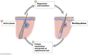
Hair Growth Cycle
Active phase: 2–5 years, hair grows 0.33 mm/day.
Resting phase: Hair loses attachment, becomes club hair, shed when follicle reactivates.
Cycle variations: Determine hair length.
Exocrine Glands of the Skin
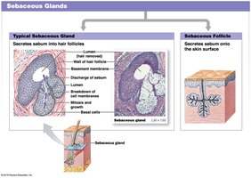
Sebaceous Glands
Holocrine glands: Discharge oily sebum onto hair and skin.
Sebum: Triglycerides, cholesterol, proteins, electrolytes; lubricates and is antimicrobial.
Sweat Glands
Apocrine: Found in axillae, nipples, pubic region; sticky, odorous secretion; influenced by hormones.
Merocrine (eccrine): Secrete directly onto skin; most numerous on palms and soles; important for thermoregulation.
Nails: Structure and Function
Nail Anatomy
Nails are thick sheets of keratinized epidermal cells that protect the tips of fingers and toes.
Nail body: Visible portion, covers nail bed.
Lunula: Pale crescent at proximal nail.
Free edge: Distal part of nail body.
Nail root: Site of nail production.
Eponychium: Cuticle, extension of stratum corneum.
Hyponychium: Thickened stratum corneum under free edge.
Clinical Module: Effects of Aging on the Skin
Age-Related Changes
Fewer melanocytes: Pale skin, increased sun sensitivity.
Drier epidermis: Decreased sebaceous activity.
Thinning epidermis: Weaker connections, more injury risk.
Reduced vitamin D3 production: Muscle weakness, brittle bones.
Diminished immune response: Fewer dendritic cells.
Thinning dermis: Fewer elastic fibers, sagging, wrinkling.
Decreased perspiration: Less active sweat glands, overheating risk.
Reduced blood supply: Cools skin, slower repair.
Fewer active follicles: Thinner, gray/white hair.
Endocrine Interactions and Vitamin D3 Production
Hormonal Regulation of Skin
Glucocorticoids: Loosen intercellular connections, reduce barrier effectiveness.
Thyroid hormones: Maintain blood flow.
Sex hormones: Increase epidermal thickness, accelerate healing.
Growth factors: Stimulate cell growth and division.
Growth hormone: Stimulates fibroblast activity, collagen production, wound repair.
Vitamin D3 Synthesis
UV radiation: Converts steroid in epidermal cells to cholecalciferol (vitamin D3).
Liver and kidneys: Convert cholecalciferol to calcitriol, enabling calcium and phosphate absorption.
Dietary sources: Fish, fish oils, shellfish, fortified foods.
Deficiency: Leads to rickets in children, decreased bone density in elderly.
Skin Repair and Regeneration
Phases of Skin Regeneration
The skin can often repair itself after injury through four distinct phases.
Inflammation phase: Bleeding and mast cell activation; inflammation causes swelling, redness, heat, pain.
Migration phase: Blood clot forms; basal cells migrate; macrophages remove debris; granulation tissue forms.
Proliferation phase: Epidermal cells migrate; phagocytic activity nearly complete; fibroblasts form collagen.
Scarring phase: Scab shed; epidermis complete; scar tissue forms.
Keloids
Keloids: Raised, thickened scar tissue that grows beyond injury site; common on upper back, shoulders, chest, earlobes.
