 Back
Integumentary System: Structure, Function, and Clinical Aspects
Study Guide - Smart Notes
 Tailored notes based on your materials, expanded with key definitions, examples, and context.
Integumentary System
Overview and Functions
The integumentary system is composed of the skin and its accessory structures, serving as the body's primary barrier against the external environment. It performs several essential functions necessary for maintaining homeostasis and overall health.
Protection: Shields underlying tissues and organs from mechanical damage, pathogens, and chemical exposure.
Excretion: Removes salts, water, and metabolic wastes through sweat.
Temperature Regulation: Maintains body temperature via sweat production and blood flow regulation.
Vitamin D3 Synthesis: Produces vitamin D3 (cholecalciferol) when exposed to UV light, which is converted to calcitriol for calcium absorption.
Storage: Stores lipids in adipocytes within the dermis and subcutaneous layer.
Sensory Reception: Contains receptors for touch, pressure, pain, and temperature.
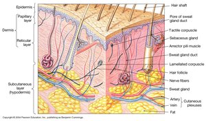
Structure of the Skin
Major Layers of the Skin
The skin consists of two main layers and an underlying subcutaneous layer:
Epidermis: The outermost layer, composed of stratified squamous epithelium; avascular.
Dermis: Lies beneath the epidermis, made of dense irregular connective tissue; highly vascularized.
Subcutaneous Layer (Hypodermis): Not technically part of the skin; consists of areolar and adipose tissue, providing insulation and energy storage.
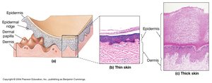
Types of Skin
Thick Skin: Found on palms and soles; contains five epidermal layers.
Thin Skin: Covers most of the body; contains four epidermal layers.
Epidermal Layers
Strata of the Epidermis
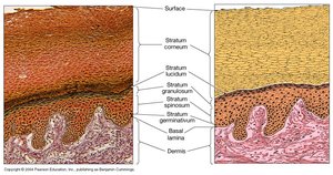
The epidermis is organized into distinct layers (strata), each with specialized cells and functions:
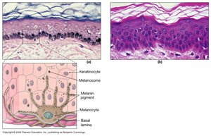
Stratum Germinativum (Basale): Deepest layer; contains stem cells (basal cells), melanocytes (produce melanin), and Merkel cells (touch receptors). Responsible for continuous cell division and attachment to the basal lamina.
Stratum Spinosum: 8–10 layers of keratinocytes; contains Langerhans cells (immune defense).
Stratum Granulosum: 3–5 layers; cells stop dividing and begin producing keratin.
Stratum Lucidum: Present only in thick skin; clear, densely packed keratinized cells.
Stratum Corneum: Outermost layer; 15–30 layers of dead, keratinized cells providing a water-resistant barrier.
Keratinization and Cell Turnover
Keratinization: The process by which cells fill with keratin and move toward the surface, forming a protective layer.
It takes approximately 15–30 days for a cell to migrate from the stratum basale to the stratum corneum.
Dead cells remain on the surface for about two weeks before being shed.
Pigmentation and Skin Color
Major Pigments
Carotene: Orange/yellow pigment from diet; converted to vitamin A, important for vision and epithelial health.
Melanin: Brown to black pigment produced by melanocytes; protects against UV radiation and determines skin color. Melanin production increases with sun exposure.
Hemoglobin: Red pigment in blood; gives skin a pinkish hue when oxygenated, bluish when deoxygenated.
Clinical Correlations
Jaundice: Yellowing of skin and eyes due to bilirubin accumulation from liver dysfunction.
Albinism: Genetic condition resulting in lack of melanin production.
Dermis and Subcutaneous Layer
Dermal Structure
Papillary Layer: Superficial; areolar tissue with capillaries and sensory neurons.
Reticular Layer: Deep; dense irregular connective tissue with collagen and elastic fibers, blood vessels, and accessory structures.
The dermis supports the epidermis and houses hair follicles, glands, blood vessels, and nerves.
Subcutaneous Layer (Hypodermis)
Composed of areolar and adipose tissue.
Functions in insulation, energy storage, and shock absorption.
Common site for subcutaneous injections.
Accessory Structures
Hair
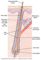
Hair is produced by hair follicles and serves protective and sensory functions.
Hair Shaft: Visible part above the skin surface; composed of dead, keratinized cells.
Hair Root: Anchors hair within the skin.
Hair Papilla: Connective tissue at the base of the follicle containing capillaries and nerves.
Arrector Pili Muscle: Smooth muscle that causes hair to stand up (goosebumps).
Hair Color: Determined by melanin; decreases with age, leading to gray hair.
Nails
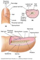
Nails are protective coverings on the dorsal surfaces of fingers and toes.
Nail Body: Visible portion covering the nail bed.
Nail Root: Site of nail growth.
Eponychium (Cuticle): Extension of the stratum corneum over the nail root.
Lunula: Pale, crescent-shaped area near the root due to obscured blood vessels.
Glands
Sebaceous (Oil) Glands: Secrete sebum to lubricate hair and skin; holocrine secretion.
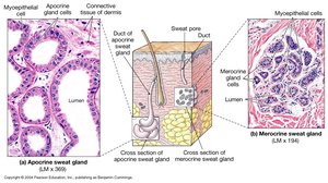
Sudoriferous (Sweat) Glands: Two types:
Apocrine: Found in armpits and groin; secrete into hair follicles; produce odor when metabolized by bacteria.
Merocrine (Eccrine): Widely distributed; secrete directly onto skin surface; involved in thermoregulation and excretion.
Mammary Glands: Modified sweat glands producing milk.
Ceruminous Glands: Modified sweat glands in the ear canal producing cerumen (earwax).
Skin Repair and Aging
Repair of Skin Injury
Skin repair involves a series of coordinated steps:
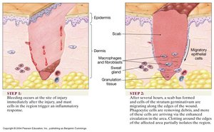
Hemostasis: Bleeding occurs, clot forms, and mast cells trigger inflammation.
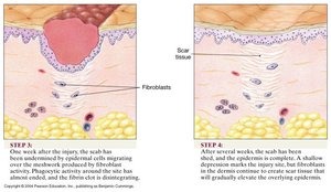
Migration: Scab forms; cells from the stratum basale migrate to cover the wound; increased blood flow brings phagocytes.
Proliferation: Epidermal cells and fibroblasts produce new tissue and scar formation begins.
Maturation: Scab is shed; fibroblasts continue to strengthen the area, sometimes resulting in a raised scar (keloid).
Aging and the Integumentary System
Epidermis thins and cell turnover decreases.
Reduced number of Langerhans cells and melanocytes.
Decreased vitamin D3 production and gland activity.
Loss of collagen and elastin leads to thinner, weaker, and more wrinkled skin.
Reduced moisture content causes dryness.
Clinical Conditions and Disorders
Common Disorders
Psoriasis: Rapid cell division in the stratum basale; leads to thick, scaly patches.
Albinism: Genetic defect preventing melanin production.
Skin Cancers:
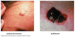
Basal Cell Carcinoma: Originates in the stratum basale; most common skin cancer.
Melanoma: Malignant tumor of melanocytes; highly dangerous.
Infections: Viral (herpes, chickenpox), bacterial (boils, leprosy), fungal (ringworm), and parasitic (scabies).
Key Terms and Definitions
Cutaneous Membrane: The skin; composed of epidermis and dermis.
Melanocytes: Cells producing melanin pigment.
Keratin: Structural protein in skin, hair, and nails.
Epidermal Ridges: Extensions of the epidermis into the dermis, increasing surface area for attachment.
Papillary Layer: Superficial dermal layer with areolar tissue.
Reticular Layer: Deep dermal layer with dense irregular connective tissue.
Elastic Fibers: Provide skin elasticity.
Collagen: Provides strength and structure to the skin.
Hair Follicle: Structure producing hair.
Arrector Pili Muscle: Muscle causing hair to stand erect.
Sebaceous Gland: Oil-producing gland associated with hair follicles.
Nail Root: Site of nail growth.
Nail Bed: Skin beneath the nail body.
Nail Fold: Skin folds on the sides of the nail.
Hyponychium: Skin under the free edge of the nail.
Eponychium: Cuticle; extension of the stratum corneum over the nail root.
Lunula: Crescent-shaped area at the nail base.
