Back
Introduction to Anatomy & Physiology: Anatomical Terms, Cells, and the Integumentary System
Study Guide - Smart Notes
 Tailored notes based on your materials, expanded with key definitions, examples, and context.
Introduction to Anatomy & Physiology
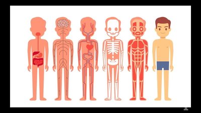
Overview of Human Body Systems
Anatomy and Physiology (A&P) is the study of the structure and function of the human body. Understanding the organization of the body and its systems is foundational for all health sciences.
Anatomy focuses on the structure of body parts and their relationships to one another.
Physiology is the study of the function of the body’s structural machinery.
Anatomical Terminology
Anatomical Position and Directional Terms
Special terminology is used in anatomy to prevent misunderstanding and to describe the location of body structures precisely. The standard anatomical position is the reference point for all directional terms.
Anatomical Position: Body erect, feet slightly apart, palms facing forward, thumbs pointing away from the body.
Directional Terms: Used to describe the position of one body part relative to another. Always based on the standard anatomical position.
Right and Left: Refer to the body being viewed, not the observer’s right and left.

Orientation and Directional Terms
Directional terms help describe the locations of structures in relation to other structures or locations in the body.
Term | Definition | Example |
|---|---|---|
Medial | Toward or at the midline of the body; on the inner side of | The heart is medial to the arm. |
Lateral | Away from the midline of the body; on the outer side of | The arms are lateral to the chest. |
Intermediate | Between a more medial and a more lateral structure | The collarbone is intermediate between the breastbone and shoulder. |

Regional Terms
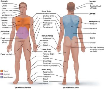
Regional terms are used to designate specific areas within major body divisions. The body is divided into two main regions: axial (head, neck, trunk) and appendicular (limbs).
Axial region: Includes the head, neck, and trunk.
Appendicular region: Includes the limbs (arms and legs).

Body Planes and Sections
Body planes are imaginary lines used to divide the body into sections for anatomical study and medical imaging.
Sagittal Plane: Divides the body vertically into right and left parts.
Midsagittal (median) plane: Cut made perfectly on the midline.
Parasagittal plane: Cut off-centered, not on the midline.
Frontal (Coronal) Plane: Divides the body vertically into anterior (front) and posterior (back) parts.
Transverse (Horizontal) Plane: Divides the body horizontally into superior (top) and inferior (bottom) parts; produces a cross-section.

Body Cavities and Membranes
Major Body Cavities
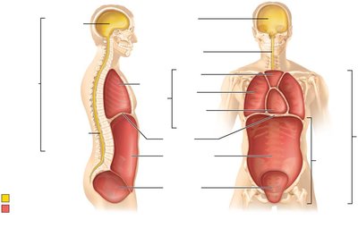
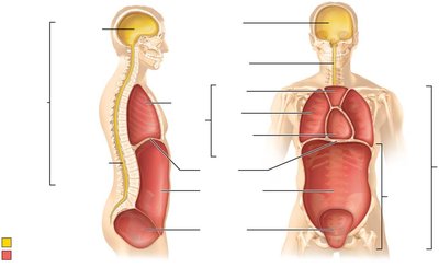
The body contains internal cavities that protect organs and allow for organ movement and expansion.
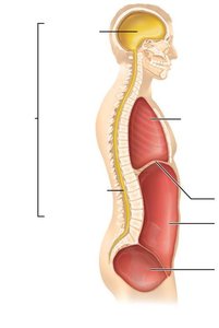
Dorsal Body Cavity: Protects the nervous system; subdivided into:
Cranial cavity: Encases the brain.
Vertebral cavity: Encases the spinal cord.
Ventral Body Cavity: Houses internal organs (viscera); subdivided into:
Thoracic cavity: Contains heart and lungs.
Abdominopelvic cavity: Contains digestive, urinary, and reproductive organs.

Serous Membranes of the Ventral Body Cavity
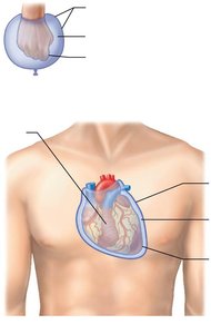
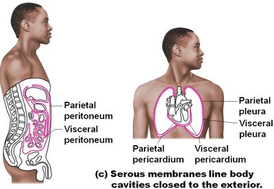
Serous membranes (serosae) are thin, double-layered membranes that cover surfaces in the ventral body cavity.
Parietal serosa: Lines internal body cavity walls.
Visceral serosa: Covers internal organs (viscera).
The two layers are separated by a slit-like cavity filled with serous fluid, which reduces friction.
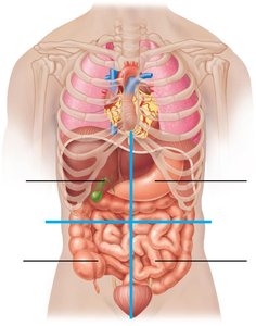
Abdominopelvic Quadrants and Regions
The abdominopelvic cavity is divided into quadrants and regions for clinical and anatomical reference.
Quadrants: Right upper (RUQ), Left upper (LUQ), Right lower (RLQ), Left lower (LLQ).
Regions: Nine regions resembling a tic-tac-toe grid, including right/left hypochondriac, epigastric, umbilical, lumbar, iliac (inguinal), and hypogastric regions.

Cells: The Living Units
Structure of the Generalized Cell
Cells are the basic structural and functional units of life. A generalized cell contains several key organelles, each with specific functions.
Nucleus: Contains genetic material (DNA) and controls cellular activities.
Plasma membrane: Encloses the cell, controlling the movement of substances in and out.
Cytoplasm: Jelly-like fluid where organelles are suspended.
Mitochondria: Produce ATP, the cell’s energy currency.
Endoplasmic reticulum (rough and smooth): Synthesizes proteins and lipids.
Golgi apparatus: Modifies, sorts, and packages proteins and lipids for secretion or use within the cell.
Ribosomes: Sites of protein synthesis.
Cytoskeleton: Maintains cell shape and assists in movement.

The Integumentary System
Overview and Functions
The integumentary system consists of the skin and its derivatives (hair, nails, sweat glands, and sebaceous glands). It serves as the body’s first line of defense and plays a role in protection, sensation, temperature regulation, and vitamin D synthesis.
Skin is the largest organ of the body.
Hair and nails are protective structures.
Sweat glands help regulate body temperature.
Sebaceous (oil) glands secrete sebum to lubricate and waterproof the skin and hair.

Layers of the Skin
The skin is composed of three main layers, each with distinct structures and functions.
Epidermis: The outermost, avascular layer made of keratinized stratified squamous epithelium.
Dermis: The middle layer, primarily dense irregular connective tissue, containing blood vessels, nerves, and accessory structures.
Hypodermis (subcutaneous layer): Not technically part of the skin, but anchors skin to underlying tissues and provides insulation and shock absorption (mainly adipose tissue).

Structures of the Skin
Several accessory structures are associated with the skin, contributing to its protective and regulatory functions.
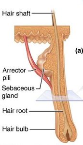
Arrector pili muscle: Small band of smooth muscle attached to hair follicles; responsible for "goose bumps."
Sebaceous glands: Secrete sebum into hair follicles for lubrication.
Sweat glands (sudoriferous glands): Produce sweat for thermoregulation.

Structure of Hair and Hair Follicle
Hair is composed of dead, keratinized cells and is produced by hair follicles. It consists of three main parts and is surrounded by specialized structures.
Hair shaft: Visible part above the skin surface; composed of medulla (core), cortex (middle), and cuticle (outer layer).
Hair follicle: Extends from the epidermis into the dermis; surrounds the hair root.
Hair papilla: Dermal tissue at the base of the follicle, supplying nutrients for hair growth.

Nails
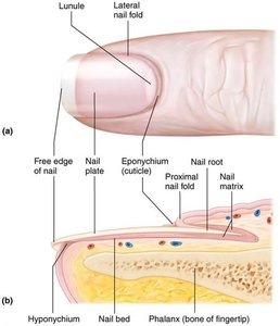
Nails are protective coverings on the dorsal surface of the distal fingers and toes. They are composed of hard keratin and have several distinct parts.
Free edge: The part that extends beyond the finger or toe.
Nail plate: The visible attached portion.
Nail root: Embedded in the skin.
Nail bed: The epidermis beneath the nail plate.
Nail matrix: Thickened portion responsible for nail growth.
Nail folds: Skin folds overlapping the nail borders.
Eponychium (cuticle): Proximal nail fold projecting onto the nail body.
Hyponychium: Area under the free edge that accumulates dirt.
Lunule: White crescent-shaped area at the base of the nail, representing the thickened nail matrix.
Abnormal color or shape can indicate disease.