 Back
BackIntroduction to Anatomy and Physiology: Anatomical Terms, Body Planes, and Cavities
Study Guide - Smart Notes
 Tailored notes based on your materials, expanded with key definitions, examples, and context.
Introduction to Anatomical Terms
Anatomical Position and Body Landmarks
The anatomical position is a standardized stance used as a reference in describing the location and relation of body parts. In this position, the body stands upright, facing forward, with arms at the sides and palms facing forward. Anatomical landmarks are specific regions of the body used to identify locations for study and clinical reference.
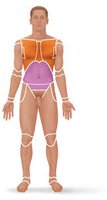
Anterior (ventral) body landmarks: Abdominal, Axillary, Brachial, Carpal, Cervical, Femoral, Frontal, Nasal, Pelvic, Sternal, Tarsal, Thoracic
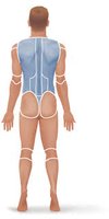
Posterior (dorsal) body landmarks: Calcaneal, Gluteal, Lumbar, Occipital, Sacral
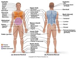
Example: The brachial region refers to the upper arm, while the femoral region refers to the thigh.
Body Regions and Landmarks Practice
Learning to identify body regions is essential for accurate communication in anatomy and clinical settings. Practice by labeling diagrams and associating terms with their locations.
Body Orientation and Direction
Directional Terms
Directional terms describe the positions of structures relative to other structures or locations in the body. These terms are essential for clear anatomical communication.
Superior/Inferior: Toward the head/away from the head
Anterior (ventral)/Posterior (dorsal): Toward the front/toward the back
Medial/Lateral: Toward the midline/away from the midline
Proximal/Distal: Closer to the point of attachment/farther from the point of attachment
Superficial/Deep: Toward or at the body surface/away from the body surface
Ventral/Dorsal: Synonymous with anterior/posterior in humans
Axial/Appendicular: Relating to the head, neck, and trunk/relating to limbs and their attachments
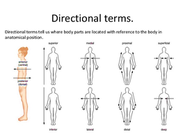
Example: The heart is medial to the lungs, and the wrist is distal to the elbow.
Body Planes and Sections
Major Body Planes
Body planes are imaginary lines used to divide the body into sections for anatomical study and medical imaging. The three primary planes are:
Sagittal Plane: Divides the body into right and left parts. The midsagittal (median) plane divides the body into equal right and left halves.
Frontal (Coronal) Plane: Divides the body into anterior (front) and posterior (back) parts.
Transverse (Horizontal) Plane: Divides the body into superior (upper) and inferior (lower) parts.
Oblique Plane: Passes through the body at an angle between the horizontal and vertical planes.
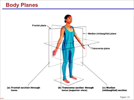
Example: A CT scan often uses the transverse plane to create cross-sectional images of the body.
Body Cavities
Major Body Cavities and Subdivisions
The human body contains several major cavities that house and protect vital organs. These cavities are divided into dorsal and ventral groups, each with further subdivisions.
Dorsal Cavity: Contains the cranial cavity (brain) and vertebral (spinal) cavity (spinal cord).
Ventral Cavity: Contains the thoracic cavity (heart and lungs) and abdominopelvic cavity (digestive, urinary, and reproductive organs).
Thoracic Cavity: Subdivided into pleural cavities (lungs), pericardial cavity (heart), and mediastinum (central compartment).
Abdominopelvic Cavity: Subdivided into abdominal cavity (digestive organs) and pelvic cavity (urinary bladder, reproductive organs, rectum).
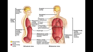
Example: The spinal cord is located within the vertebral cavity, a subdivision of the dorsal cavity.
Axial and Appendicular Divisions
Axial vs. Appendicular Skeleton
The body is structurally divided into the axial and appendicular regions. The axial region includes the head, neck, and trunk, while the appendicular region consists of the limbs and their attachments to the axis.
Axial: Skull, vertebral column, rib cage
Appendicular: Limbs (arms and legs), pelvic and pectoral girdles
Example: The femur is part of the appendicular skeleton, while the sternum is part of the axial skeleton.
Additional info: The distinction between axial and appendicular regions is fundamental for understanding skeletal anatomy and movement.
