 Back
BackIntroduction to Anatomy & Physiology: Foundational Concepts and Terminology
Study Guide - Smart Notes
 Tailored notes based on your materials, expanded with key definitions, examples, and context.
Introduction to Anatomy & Physiology
What is Anatomy and Physiology?
Anatomy and Physiology (A&P) is the study of the structure and function of the human body. Anatomy focuses on the physical structures of the body, while physiology examines how those structures function and interact.
Anatomy: The study of the body’s structures (e.g., bones, muscles, organs).
Physiology: The study of the body’s functions (e.g., how the heart pumps blood, how muscles contract).
Principle of Complementarity: Structure and function are closely related; understanding one helps explain the other.
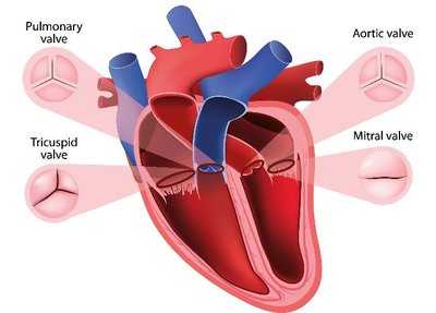
Example: The heart’s structure (chambers and valves) allows it to function as a pump for blood circulation.
Relationship Between Anatomy and Physiology
To understand why an organ is shaped a certain way, you must understand what it does. Conversely, to understand how an organ performs its job, you must understand its structure.
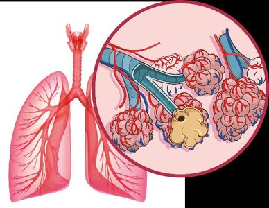
Example: The lungs have thin walls and many alveoli to maximize gas exchange.
Levels of Organization
Hierarchy of Structural Organization
The human body is organized into a hierarchy of levels, each building on the previous one:
Atomic and Molecular Level: Atoms and molecules form the chemical basis of life.
Macromolecule Level: Large molecules such as proteins and DNA.
Cellular Level: Cells are the basic units of life.
Tissue Level: Groups of similar cells performing a common function.
Organ Level: Structures composed of two or more tissue types.
Organ System Level: Groups of organs working together for a common purpose.
Organism Level: The complete living being.
Additional info: Changes at one level can affect other levels, demonstrating the interconnectedness of the body’s organization.
Variation in Anatomy and Physiology
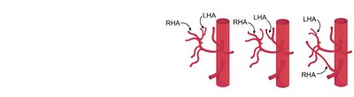
Anatomical Variation
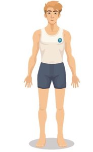
There is significant variation in human anatomy. Textbooks often present a ‘reference body’ for simplicity, but real individuals may differ in size, shape, and internal arrangement of organs.
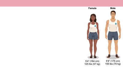
Reference Body: Typically a healthy young adult of average size (female: 5’4”, 125 lbs; male: 5’9”, 155 lbs).
Normal Variation: Most variations do not affect function, but some can lead to medical errors if not recognized.
Introduction to Organ Systems
Overview of Organ Systems
The body is organized into organ systems, each with specific functions. These systems are highly integrated and often work together.
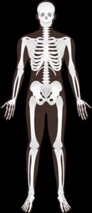
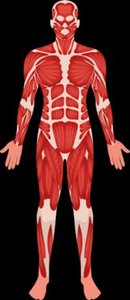
Protection, Structure, & Support: Integumentary, Skeletal, Muscular systems
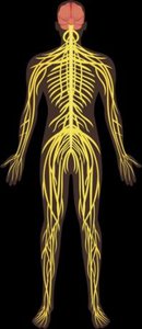
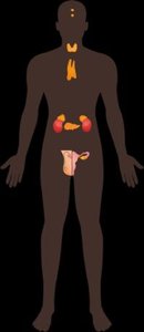
Communication & Integration: Nervous, Endocrine systems
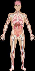
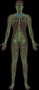
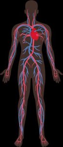
Transport & Immunity: Cardiovascular (Circulatory), Lymphatic systems
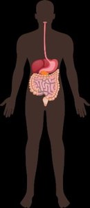
Nutrient, Gas, & Waste Exchange: Respiratory, Digestive, Urinary systems
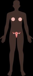
Reproduction: Male and Female Reproductive systems

Homeostasis
Definition and Importance
Homeostasis is the maintenance of a stable internal environment. The body regulates variables such as temperature, pH, and blood glucose within narrow ranges to sustain life.
Dynamic Equilibrium: The body is always adjusting to maintain balance, not a fixed state.
Pathology: Failure to maintain homeostasis leads to disease.
Examples of Homeostatic Variables
Variable | Normal Range | Pathology (Out of Range) |
|---|---|---|
Blood pH | 7.35–7.45 | Acidosis or alkalosis |
Internal Body Temperature | 36–37.5°C (97–99.5°F) | Hypothermia or hyperthermia |
Blood Glucose | 70–90 mg/dL (fasting) | Hypoglycemia or hyperglycemia (diabetes) |
Feedback Loops
Types of Feedback Loops
Homeostasis is maintained through feedback loops, which can be negative or positive.
Negative Feedback: Opposes the original stimulus, returning the system to a set point. Most common type.
Positive Feedback: Amplifies the original stimulus, moving the system further from the set point. Less common, used in specific situations (e.g., blood clotting, childbirth).

Negative Feedback Example: Thermoregulation
When body temperature rises, sweat glands produce sweat and blood vessels dilate to cool the body. When temperature falls, muscles shiver and blood vessels constrict to conserve heat.


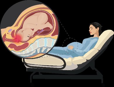
Positive Feedback Example: Childbirth
During labor, pressure on the cervix stimulates the release of oxytocin, which increases uterine contractions, leading to more pressure and more oxytocin until delivery occurs.
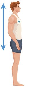
Anatomical Position and Directional Terms
Anatomical Position
The anatomical position is a standard reference for describing body parts and positions. The body stands upright, facing forward, feet shoulder-width apart, arms at the sides, and palms facing forward. Left and right refer to the subject’s left and right.
Directional Terms
Superior (Cranial): Toward the head
Inferior (Caudal): Toward the feet
Anterior (Ventral): Toward the front
Posterior (Dorsal): Toward the back
Medial: Toward the midline
Lateral: Away from the midline
Proximal: Closer to the point of attachment (limbs)
Distal: Farther from the point of attachment (limbs)
Superficial: Closer to the surface
Deep: Further from the surface
Anatomical Planes and Sections
Major Anatomical Planes
Frontal (Coronal) Plane: Divides the body into anterior and posterior parts.
Sagittal Plane: Divides the body into right and left parts. Midsagittal is exactly at the midline; parasagittal is off-center.
Transverse (Horizontal) Plane: Divides the body into superior and inferior parts.
Oblique Plane: Divides the body at an angle.
Body Cavities and Serous Membranes
Major Body Cavities
Anterior (Ventral) Cavity: Houses most organs; divided into thoracic and abdominopelvic cavities by the diaphragm.
Posterior (Dorsal) Cavity: Contains the brain and spinal cord.
Serous Membranes
Serous membranes are double-layered membranes that line body cavities and cover organs. They secrete serous fluid to reduce friction.
Visceral Layer: Attached to the organ.
Parietal Layer: Attached to the cavity wall.
Serous Cavity: The space between the two layers, filled with serous fluid.
Major Serous Membranes
Pleura: Surrounds the lungs.
Pericardium: Surrounds the heart.
Peritoneum: Surrounds most abdominal organs.
Abdominopelvic Quadrants and Regions
Quadrants
The abdomen is divided into four quadrants for clinical reference:
Right Upper Quadrant (RUQ)
Left Upper Quadrant (LUQ)
Right Lower Quadrant (RLQ)
Left Lower Quadrant (LLQ)
Regions
For more precise localization, the abdomen is divided into nine regions:
Right/Left Hypochondriac
Epigastric
Right/Left Lumbar
Umbilical
Right/Left Inguinal
Hypogastric
Summary Table: Major Organ Systems and Functions
System | Main Organs | Primary Function |
|---|---|---|
Integumentary | Skin, hair, nails | Protection, temperature regulation |
Skeletal | Bones, cartilage | Support, movement, protection |
Muscular | Muscles | Movement, heat production |
Nervous | Brain, spinal cord, nerves | Rapid communication, control |
Endocrine | Glands (pituitary, thyroid, etc.) | Hormonal regulation |
Cardiovascular | Heart, blood vessels | Transport of nutrients, gases, wastes |
Lymphatic | Lymph nodes, vessels, spleen | Immunity, fluid balance |
Respiratory | Lungs, trachea | Gas exchange |
Digestive | Stomach, intestines, liver | Breakdown and absorption of food |
Urinary | Kidneys, bladder | Waste removal, water balance |
Reproductive | Ovaries/testes, uterus/penis | Production of offspring |
