 Back
CH 1: Introduction to Anatomy and Physiology: Levels of Organization, Terminology, and Homeostasis
Study Guide - Smart Notes
 Tailored notes based on your materials, expanded with key definitions, examples, and context.
Ch 1: Levels of Organization in the Human Body
The Six Major Levels
The human body is organized into hierarchical levels, each with increasing complexity. Understanding these levels is fundamental to studying anatomy and physiology.
Chemical Level: Atoms and molecules form the basis of all matter. The arrangement and structure of molecules, such as proteins, determine their function in the body.
Cellular Level: Cells are the smallest living units. Organelles within cells perform specialized functions, such as energy production for muscle contraction.
Tissue Level: Tissues are groups of similar cells working together. For example, cardiac muscle tissue is formed by heart muscle cells and other supporting cells.
Organ Level: Organs consist of two or more tissues working together for specific functions, such as the heart's pumping action.
Organ System Level: Organ systems are groups of organs that perform coordinated functions. The cardiovascular system includes the heart, blood, and blood vessels.
Organism Level: The highest level, representing the complete living human, where all systems work together to maintain life.
Additional info: Disruption at any level can affect the entire organism, as seen in cases of massive blood loss affecting all systems.
Medical Terminology in Anatomy and Physiology
Origins and Importance
Medical terminology is essential for precise communication in anatomy and physiology. Most terms are derived from Greek and Latin roots, and standardized naming avoids confusion.
Word Roots: The core meaning of a term (e.g., path- for disease).
Prefixes: Added to the beginning to modify meaning (e.g., peri- for around).
Suffixes: Added to the end to form new terms (e.g., -ology for study of).
Combining Forms: Used to build complex terms.
Standardized texts such as Terminologia Anatomica ensure consistency in naming structures.
Anatomical Terms and Body Regions
Surface Anatomy and Anatomical Landmarks
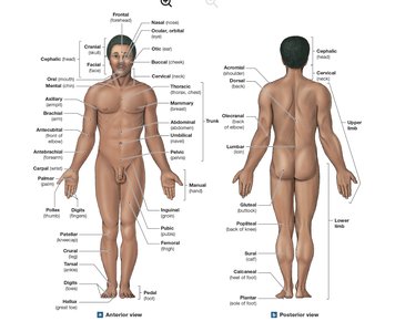
Surface anatomy involves identifying structures on or near the body surface using landmarks and regions. Anatomical position is the standard reference: standing, hands at sides, palms forward, feet together.
Anterior View: Front of the body.
Posterior View: Back of the body.
Supine: Lying face up.
Prone: Lying face down.
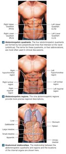
Abdominopelvic Quadrants and Regions
The abdomen and pelvis are mapped using quadrants and regions for clinical and anatomical reference.
Quadrants: Four areas (RUQ, RLQ, LUQ, LLQ) used to localize symptoms.
Regions: Nine more precise areas for internal organ location.
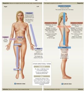
Anatomical Directions
Directional terms describe the location of structures relative to each other.
Anterior/Ventral: Front/belly side.
Posterior/Dorsal: Back side.
Superior: Above.
Inferior: Below.
Medial: Toward the midline.
Lateral: Away from the midline.
Proximal: Closer to the point of attachment.
Distal: Farther from the point of attachment.
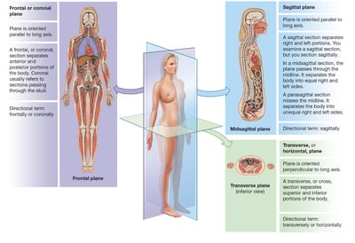
Sectional Anatomy
Sectional anatomy uses planes to describe slices through the body, aiding in understanding internal relationships.
Frontal (Coronal) Plane: Divides into anterior and posterior portions.
Sagittal Plane: Divides into left and right portions (midsagittal or parasagittal).
Transverse (Horizontal) Plane: Divides into superior and inferior portions.
Body Cavities of the Trunk
Major Cavities and Their Functions
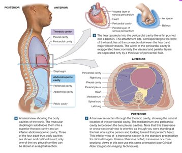
The trunk contains three major regions: thoracic, abdominal, and pelvic. True body cavities are closed, fluid-filled, and lined by serous membranes.
Thoracic Cavity: Contains pleural cavities (lungs), pericardial cavity (heart), and mediastinum.
Abdominopelvic Cavity: Contains abdominal and pelvic organs, lined by the peritoneum.
Functions: Protection from shocks, and allowing organ shape/size changes.
Serous Membranes: Visceral serosa covers organs; parietal serosa lines cavity walls.
Homeostasis and Physiological Regulation
Definition and Mechanisms
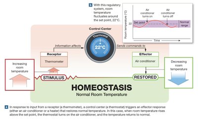
Homeostasis is the maintenance of a stable internal environment through continuous physiological processes. It is essential for survival.
Autoregulation: Local adjustment by cells/tissues/organs in response to environmental changes.
Extrinsic Regulation: Adjustment by nervous or endocrine systems, affecting multiple systems.
Homeostatic regulation involves three components:
Receptor: Detects changes.
Control Center: Processes information and sends commands.
Effector: Responds to restore balance.
Negative and Positive Feedback in Homeostasis
Negative Feedback
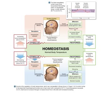
Negative feedback opposes deviations from normal, minimizing change. Example: thermoregulation, where the hypothalamus adjusts blood flow and sweat production to maintain body temperature.
Set Point: Desired value range (e.g., 37°C for body temperature).
Effectors: Blood vessels and sweat glands adjust to restore normal temperature.
Positive Feedback
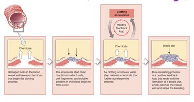
Positive feedback amplifies changes, producing rapid responses. Example: blood clotting, where chemicals released by damaged cells accelerate clot formation until bleeding stops.
Positive Feedback Loop: Response enhances the original stimulus.
Clinical Examples: Blood clotting, labor and delivery.
Systems Integration and Dynamic Equilibrium
Organ Systems and Homeostasis
Multiple organ systems coordinate to maintain homeostasis and dynamic equilibrium. Adjustments in one system affect others, requiring integrated regulation.
Equilibrium: Balance between opposing processes (e.g., heat loss and production).
Dynamic Equilibrium: Continuous adaptation to changing conditions.
Table: Roles of Organ Systems in Homeostatic Regulation
Internal Stimulus | Primary Organ Systems Involved | Functions of the Organ Systems |
|---|---|---|
Body temperature | Integumentary, muscular, cardiovascular, nervous | Heat loss, heat production, heat distribution, coordination |
Body fluid composition | Digestive, cardiovascular, urinary, respiratory, lymphatic | Nutrient absorption, storage, release; mineral storage; gas exchange; removal of wastes and pathogens |
Body fluid volume | Urinary, digestive, cardiovascular, lymphatic | Elimination/conservation of water; distribution of water |
Waste concentration | Urinary, digestive | Excretion of wastes |
Blood pressure | Cardiovascular, nervous, endocrine | Transport of nutrients/wastes; regulation of blood vessel diameter and blood pressure |
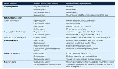
Additional info: Homeostatic set points vary among individuals due to genetic, age, sex, health, and environmental factors. Illness or disease results when homeostatic mechanisms fail to compensate for severe disruptions.
