 Back
BackIntroduction to Anatomy & Physiology: Organization, Orientation, and Homeostasis
Study Guide - Smart Notes
 Tailored notes based on your materials, expanded with key definitions, examples, and context.
Science of Anatomy & Physiology
Definition and Scope
Anatomy and Physiology (A&P) are foundational sciences for understanding the structure and function of the human body. Anatomy is the study of body structure and the relationships among structures, while Physiology focuses on the functions of these structures.
Anatomy: Structure and relationships among body parts.
Physiology: Function of body parts and systems.
Subdivisions of Anatomy
Surface Anatomy: Study of external features and markings.
Gross (Macroscopic) Anatomy: Study of structures visible without a microscope.
Systemic Anatomy: Study of specific body systems (e.g., nervous, respiratory).
Regional Anatomy: Study of specific regions (e.g., head, chest).
Radiographic Anatomy: Study using imaging techniques (e.g., x-rays).
Developmental Anatomy: Study of development from fertilization to adulthood.
Embryology: Study of development from fertilization to the eighth week in utero.
Histology: Microscopic study of tissues.
Cytology: Microscopic and chemical study of cells.
Pathology: Study of structural changes associated with disease.
Subdivisions of Physiology
Cell Physiology: Functions of cells.
Systems Physiology: Operation of organ systems.
Pathophysiology: Functional changes due to disease and aging.
Exercise Physiology: Changes during muscular activity.
Neurophysiology: Function of nerve cells.
Endocrinology: Hormones and their control of body functions.
Cardiovascular Physiology: Functions of heart and blood vessels.
Immunology: Body defense mechanisms.
Respiratory Physiology: Functions of air passageways and lungs.
Renal Physiology: Functions of kidneys.
Reproductive Physiology: Functions of reproductive structures.
Levels of Organization
Hierarchical Structure
The human body is organized into several levels, each building upon the previous:
Chemical Level: Atoms and molecules essential for life (e.g., C, H, O, N, Ca, K, Na).
Cellular Level: Fundamental units of life.
Tissue Level: Groups of cells performing the same function.
Organ Level: Two or more tissues working together.
System Level: Related organs with a common function.
Organismic Level: All body parts functioning together.
Organ Systems
Overview of Major Systems
The body consists of eleven major organ systems, each with specific components and functions:
Integumentary System: Skin, hair, nails; regulates temperature, protects, produces vitamin D.
Skeletal System: Bones, cartilages, joints; supports, protects, stores minerals, produces blood cells.
Muscular System: Skeletal muscles; movement, posture, heat production.
Nervous System: Brain, spinal cord, nerves, sense organs; regulates activities via nerve impulses.
Endocrine System: Hormone-producing glands; regulates activities via hormones.
Cardiovascular System: Heart, blood, vessels; transports nutrients, gases, wastes, regulates temperature.
Lymphatic & Immune System: Lymph, vessels, spleen, thymus, nodes; returns proteins, defends against disease.
Respiratory System: Lungs, passageways; supplies oxygen, removes CO2, regulates acid-base balance.
Digestive System: GI tract, glands; breaks down and absorbs food, eliminates wastes.
Urinary System: Kidneys, bladder, ureters, urethra; regulates blood composition, eliminates wastes.
Reproductive System: Testes, ovaries, ducts; produces gametes, enables reproduction.
Life Processes
Characteristics of Living Organisms
To be considered alive, organisms must exhibit several key processes:
Metabolism: All chemical reactions in the body.
Catabolism: Breakdown of molecules for energy.
Anabolism: Building of structural and functional components.
Responsiveness: Ability to detect and respond to changes.
Movement: Motion of body, organs, cells, or organelles.
Growth: Increase in size and complexity.
Differentiation: Cells become specialized.
Reproduction: Formation of new cells or individuals.
Homeostasis
Definition and Importance
Homeostasis is the maintenance of a stable internal environment within physiological limits. It is essential for cell survival and overall health.
Extracellular Fluid (ECF): Fluid outside cells (interstitial fluid, plasma).
Intracellular Fluid (ICF): Fluid within cells.
Optimal concentrations of gases, nutrients, ions, water, temperature, and volume are required.
Regulation of Homeostasis
Homeostasis is regulated by the nervous and endocrine systems:
Nervous System: Detects changes and sends nerve impulses to counteract stress.
Endocrine System: Secretes hormones into the blood; slower response than nerves.
Feedback Systems
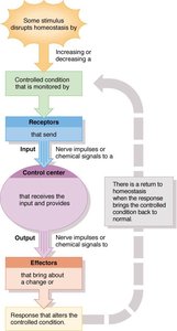
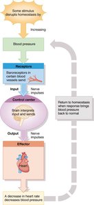
Feedback systems monitor and regulate internal conditions:
Receptor: Detects changes.
Control Center: Receives input and determines response.
Effector: Produces the response.
Negative Feedback: Response reverses the original stimulus.
Positive Feedback: Response enhances the original stimulus.
Anatomical Position and Orientation
Anatomical Position
All descriptions of the human body assume the anatomical position: upright, facing forward, feet flat, arms at sides, palms forward.
Anatomical Landmarks
Specific regions and landmarks are used to describe locations on the body:
Cephalic: Head
Cranial: Skull
Facial: Face
Frontal: Forehead
Orbital: Eye
Otic: Ear
Buccal: Cheek
Nasal: Nose
Oral: Mouth
Mental: Chin
Cervical: Neck
Thoracic: Chest
Axillary: Armpit
Brachial: Arm
Antecubital: Front of elbow
Antebrachial: Forearm
Carpal: Wrist
Metacarpal: Palm
Digital/Phalangeal: Fingers
Olecranal: Back of elbow
Acromial: Shoulder
Umbilical: Belly button
Coxal: Hip
Inguinal: Groin
Dorsal: Back
Manual: Hand
Volar: Palm
Femoral: Thigh
Patellar: Front of knee
Tarsal: Ankle
Pedal: Foot
Gluteal: Buttock
Popliteal: Back of knee
Sural: Calf
Plantar: Sole of foot
Calcaneal: Heel
Hallux: Big toe
Pollex: Thumb
Digital/Phalangeal: Toes
Regional Names and Abdominopelvic Regions
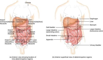
The abdomen is divided into regions for clinical and anatomical reference:
Hypochondrial
Epigastric
Umbilical
Hypogastric
Lumbar
Iliac
Abdominal Quadrants
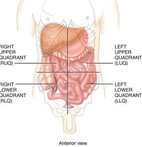
The abdomen is also divided into four quadrants for clinical assessment:
Right Upper Quadrant (RUQ)
Left Upper Quadrant (LUQ)
Right Lower Quadrant (RLQ)
Left Lower Quadrant (LLQ)
Directional Terms
Standard Anatomical Directions
Directional terms are used to describe the location of structures relative to others:
Superior: Toward the head
Inferior: Away from the head
Anterior (Ventral): Front of the body
Posterior (Dorsal): Back of the body
Medial: Closer to the midline
Lateral: Farther from the midline
Intermediate: Between two structures
Ipsilateral: Same side
Contralateral: Opposite side
Proximal: Closer to attachment or origin
Distal: Farther from attachment or origin
Superficial: Toward or on the surface
Deep: Away from the surface
Parietal: Pertaining to the outer wall of a cavity
Visceral: Pertaining to the covering of an organ
Body Cavities
Major Cavities and Their Contents
The body contains several cavities that house organs:
Dorsal Cavity: Near the back; includes cranial cavity (brain) and spinal canal (spinal cord).
Ventral Cavity: Front aspect; includes thoracic cavity (lungs, heart) and abdominopelvic cavity (digestive, urinary, reproductive organs).
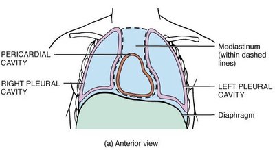
Thoracic Cavity:
Pleural Cavities: Lungs
Pericardial Cavity: Heart
Mediastinum: Central region containing heart, vessels, trachea, esophagus
Planes of the Body
Standard Anatomical Planes
Sagittal Plane: Divides body into right and left sides.
Mid-sagittal: Equal sides
Parasagittal: Unequal sides
Frontal (Coronal) Plane: Divides body into anterior and posterior portions.
Transverse Plane: Divides body into superior and inferior portions.
Oblique Plane: Passes through at an angle.
Medical Imaging
Techniques for Studying Anatomy
Conventional Radiography: X-rays for 2-D images; structures overlap.
Computed Tomography (CT): Cross-sectional, 3-D images; useful for tumors, stones, infection.
Dynamic Spatial Reconstruction (DSR): Moving, 3-D images; measures movement, tissue damage.
Digital Subtraction Angiography (DSA): Contrast dye highlights blood vessels; useful for brain and heart.
Positron Emission Tomography (PET): Radioactive tracers measure metabolic changes and function.
Magnetic Resonance Imaging (MRI): Radio waves and magnets for 2-D/3-D images; detects tumors, plaques, blood flow.
Ultrasound: High-frequency sound waves; used for fetal development, abdominal organs, blood flow.
Summary Table: Levels of Organization
Level | Description | Examples |
|---|---|---|
Chemical | Atoms and molecules | Proteins, carbohydrates, fats, vitamins |
Cellular | Basic unit of life | Muscle cell, nerve cell |
Tissue | Group of similar cells | Muscle tissue, nervous tissue |
Organ | Two or more tissues | Heart, liver |
System | Related organs | Digestive system, nervous system |
Organism | All systems together | Human body |
Summary Table: Feedback System Components
Component | Function |
|---|---|
Receptor | Detects change |
Control Center | Receives input, determines response |
Effector | Produces response |
Summary Table: Directional Terms
Term | Definition |
|---|---|
Superior | Toward the head |
Inferior | Away from the head |
Anterior (Ventral) | Front of the body |
Posterior (Dorsal) | Back of the body |
Medial | Closer to midline |
Lateral | Farther from midline |
Proximal | Closer to attachment |
Distal | Farther from attachment |
Superficial | Toward surface |
Deep | Away from surface |
Summary Table: Body Cavities
Cavity | Location | Main Contents |
|---|---|---|
Cranial | Dorsal | Brain |
Spinal | Dorsal | Spinal cord |
Thoracic | Ventral | Lungs, heart |
Abdominopelvic | Ventral | Digestive, urinary, reproductive organs |
Summary Table: Medical Imaging Techniques
Technique | Main Use |
|---|---|
Radiography | 2-D images, bone structure |
CT | Cross-sectional, 3-D images |
DSR | Moving, 3-D images |
DSA | Blood vessel imaging |
PET | Metabolic and functional imaging |
MRI | Soft tissue, blood flow |
Ultrasound | Fetal, abdominal, blood flow |
