 Back
BackIntroduction to Anatomy & Physiology: Structural Organization and Cellular Components
Study Guide - Smart Notes
 Tailored notes based on your materials, expanded with key definitions, examples, and context.
Introduction to Anatomy & Physiology
Definition and Relationship of Anatomy and Physiology
Anatomy and physiology are foundational sciences in understanding the human body. Anatomy is the study of the structure of the human body, derived from the Greek word 'anatomē' meaning dissection. Physiology is the study of the functions of the human body. These disciplines are closely related, as structure often determines function.
Anatomy: Focuses on the physical structures, such as organs, tissues, and cells.
Physiology: Explores how these structures work and interact to sustain life.
Example: The anatomy of the heart (its chambers and valves) directly influences its physiological function (pumping blood).
Hierarchy of Structural Organization
Levels of Organization in the Human Body
The human body is organized into a hierarchy of structural levels, each building upon the previous. Understanding these levels is essential for studying anatomy and physiology.
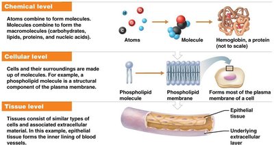
Chemical Level: Atoms combine to form molecules, which then form macromolecules (carbohydrates, lipids, proteins, nucleic acids).
Cellular Level: Molecules and macromolecules form organelles, which make up cells—the basic unit of life.
Tissue Level: Groups of similar cells and their extracellular matrix form tissues (e.g., epithelial, connective, muscle, nervous).
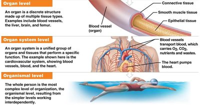
Organ Level: Organs are discrete structures made of multiple tissue types, performing specific functions (e.g., heart, liver).
Organ System Level: Groups of organs work together to perform major functions (e.g., cardiovascular system).
Organismal Level: The highest level, representing the complete living individual.
Basic Components of the Human Body
The human body is composed primarily of water, proteins, and other molecules. These components are distributed across the structural levels.
Water: 70-85% of body mass; essential for biochemical reactions.
Proteins: 10-20% of body mass; structural and functional roles.
Other: Includes inorganic salts, lipids, carbohydrates, nucleic acids.
Systemic Anatomy Approach
Systemic anatomy studies the body by examining its organ systems. Each system consists of organs and tissues working together for a common function.
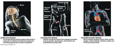
Nervous System: Controls and coordinates body activities.
Endocrine System: Regulates processes via hormones.
Cardiovascular System: Transports blood, nutrients, gases, and wastes.
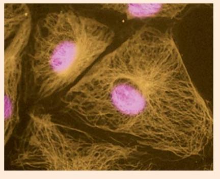
Cellular Structures and Functions
Cell Structure Overview
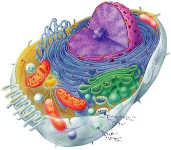
Cells are the basic units of life, containing specialized structures called organelles. Each organelle performs distinct functions necessary for cell survival.
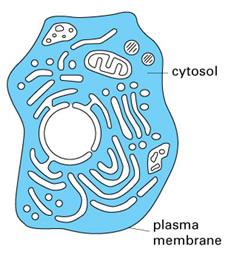
Plasma Membrane: Separates intracellular and extracellular environments; regulates entry and exit of substances; composed of a phospholipid bilayer, proteins, cholesterol, and glycocalyx.
Cytoplasm: The region between the plasma membrane and nucleus; contains cytosol, organelles, and inclusions.
Organelles: Include mitochondria, endoplasmic reticulum, Golgi apparatus, lysosomes, etc.
Plasma Membrane Structure and Function
The plasma membrane is a dynamic barrier that maintains cellular integrity and mediates communication.
Phospholipid Bilayer: Provides structural foundation.
Proteins: Integral and peripheral proteins facilitate transport and signaling.
Cholesterol: Stabilizes membrane fluidity.
Glycocalyx: Carbohydrate-rich layer involved in cell recognition.

Cytoplasm and Its Components
The cytoplasm is a semi-fluid matrix that houses organelles and inclusions.
Cytosol: Jelly-like fluid where metabolic reactions occur.
Organelles: Specialized structures performing cellular functions.
Inclusions: Temporary structures such as lipid droplets and glycosomes.
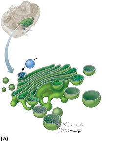
Membrane-Bound Organelles
Organelles are specialized compartments within cells, each with unique functions.
Rough Endoplasmic Reticulum (RER): Studded with ribosomes; synthesizes proteins.
Smooth Endoplasmic Reticulum (SER): Lacks ribosomes; synthesizes lipids and detoxifies chemicals.
Golgi Apparatus: Sorts, processes, and packages proteins and membranes.
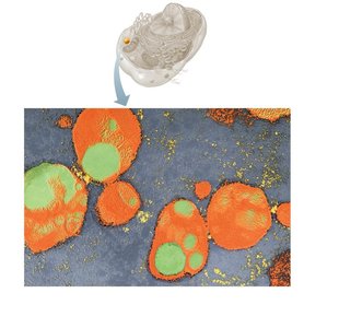
Mitochondria: Main site of energy (ATP) production.
Lysosomes: Contain digestive enzymes for breaking down cellular debris.


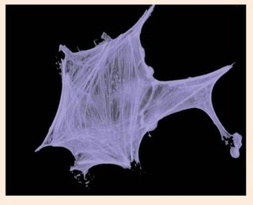
Cytoskeletal Structures and Cell Junctions
Cytoskeleton: Types and Functions
The cytoskeleton provides structural support, facilitates movement, and organizes cellular contents.
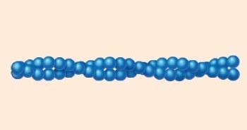
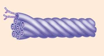
Microtubules: Largest diameter; hollow tubes of tubulin; maintain cell shape and serve as tracks for organelle movement.
Intermediate Filaments: Rope-like protein fibers; resist mechanical stress.
Microfilaments: Smallest diameter; composed of actin; involved in cell movement and shape changes.
Cells That Move
Some cells, such as muscle cells, are specialized for movement. Other examples include immune cells and sperm cells.
Cytoplasmic Inclusions
Cytoplasmic inclusions are temporary structures whose contents vary depending on cell function.
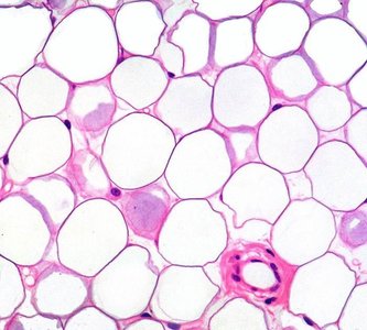
Lipid Droplets: Storage form of fats, prominent in adipocytes.
Glycosomes: Store glycogen for energy.
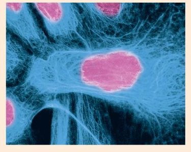
Nucleus and Its Components
The nucleus is the control center of the cell, containing genetic material and the nucleolus.
DNA: Genetic blueprint for cellular function.
Nucleolus: Produces ribosomal RNA and assembles ribosome subunits.
Nuclear Pores: Allow transport of molecules between nucleus and cytoplasm.
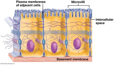
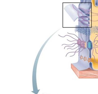
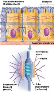
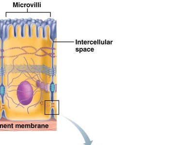
Cell Junctions: Types and Functions
Cell junctions are specialized structures that connect cells and facilitate communication.
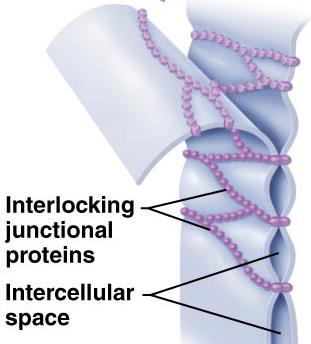
Tight Junctions: Belt-like junctions that seal adjacent cells, preventing passage of substances between them.
Adhesive Belt Junctions: Reinforce tight junctions, providing additional adhesion.
Desmosomes: Bind cells together via linker proteins and intermediate filaments, distributing mechanical stress.
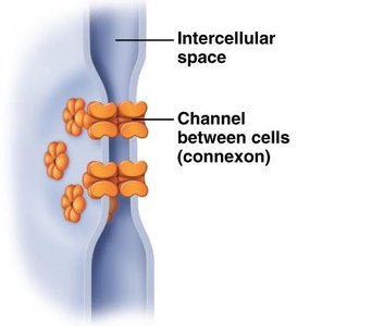
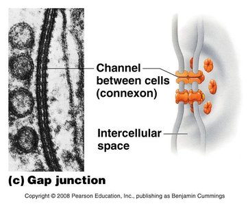
Gap Junctions: Channels that allow ions and small molecules to pass directly between cells, enabling intercellular communication.
Primary Tissue Types
Classification of Tissues
The human body contains four primary tissue types, each with distinct functions.
Epithelial Tissue: Covers surfaces and lines cavities; involved in protection, absorption, and secretion.
Connective Tissue: Supports and binds other tissues; includes bone, cartilage, blood, and adipose tissue.
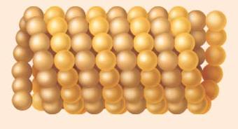
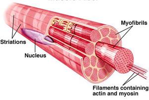
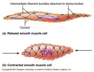
Muscle Tissue: Specialized for contraction and movement.
Nervous Tissue: Conducts electrical impulses for communication.
Summary Table: Levels of Structural Organization
Level | Description | Example |
|---|---|---|
Chemical | Atoms and molecules form macromolecules | Hemoglobin, phospholipids |
Cellular | Organelles and molecules form cells | Muscle cell, neuron |
Tissue | Groups of similar cells form tissues | Epithelial tissue, muscle tissue |
Organ | Multiple tissues form organs | Heart, liver |
Organ System | Organs work together for a function | Cardiovascular system |
Organism | Complete living individual | Human |
Key Equations and Concepts
Macromolecule Formation
Macromolecules are formed by the polymerization of monomers:
Cellular Transport Across Membranes
Diffusion is a key process for movement of substances:
Where J is the flux, D is the diffusion coefficient, and \frac{dC}{dx} is the concentration gradient.
Additional info:
Some details about cytoskeletal structures and cell junctions were inferred from standard academic sources to ensure completeness.
Table entries and equations were expanded for clarity and academic context.
