 Back
BackIntroduction to Anatomy and Physiology: Structured Study Notes
Study Guide - Smart Notes
 Tailored notes based on your materials, expanded with key definitions, examples, and context.
Introduction to Anatomy and Physiology
What is Anatomy and Physiology?
Anatomy and physiology are two closely related branches of biology that study the structure and function of the human body. Anatomy focuses on the physical structure of organisms, while physiology examines how those structures function.
Anatomy: The study of the body's structure, including organs, tissues, and systems.
Physiology: The study of the body's functions and processes.
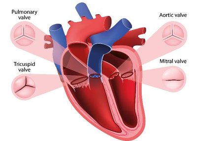
Example: The heart's anatomy includes its chambers and valves; its physiology involves how it pumps blood.
Principle of Complementarity: Structure and function are interdependent. To understand why an organ is shaped a certain way, you must understand what it does.
Anatomy & Physiology Relationship
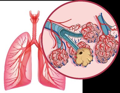
The relationship between anatomy and physiology is illustrated by the lungs:
Structure: Lungs have thin walls and many alveoli to maximize surface area.
Function: Gas exchange (O2 and CO2) between air and blood.
Principle: Function is determined by structure.
Levels of Organization
Hierarchy of Structural Organization
The human body is organized into a hierarchy of levels, each building upon the previous:
Atomic and Molecular Level: Atoms and molecules form the basis of all biological structures.
Macromolecule Level: Large molecules such as proteins and DNA.
Cellular Level: Cells are the basic unit of life.
Tissue Level: Groups of similar cells performing a common function.
Organ Level: Structures composed of multiple tissue types.
Organism Level: The complete living individual.
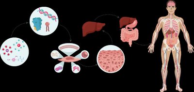
Example: Statins work at the molecular level by inhibiting an enzyme, but their effects cascade to organ and organism levels.
Variation in Anatomy and Physiology
Anatomical Variation
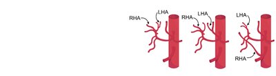
There is significant variation in human anatomy, which can impact medical procedures and understanding.
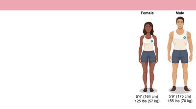
Reference Body: A standard body used for teaching and comparison, typically a healthy young adult.
Normal Variation: Most variations are harmless, but extreme variations can affect function.
Example: Variations in blood vessel branching to the liver and gall bladder.
Introduction to Organ Systems
Overview of Organ Systems
Organ systems are groups of organs that work together to perform major functions. They are often grouped by function:
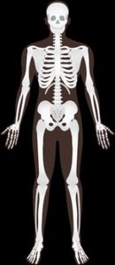
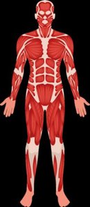
Protection, Structure, & Support: Integumentary, Skeletal, Muscular systems
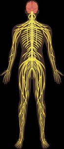
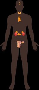
Communication & Integration: Nervous, Endocrine systems
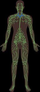
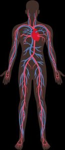
Transport & Immunity: Cardiovascular, Lymphatic systems
Nutrient, Gas, & Waste Exchange: Respiratory, Digestive, Urinary systems
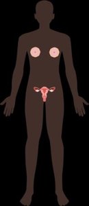
Reproduction: Male and Female Reproductive systems

Communication and Integration
Nervous System: Fast communication using electrical signals.
Endocrine System: Slow, whole-body coordination using hormones.
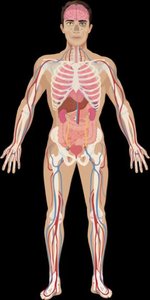
Transport and Immunity
Cardiovascular System: Transports materials through the body.
Lymphatic System: Provides immunity and transports lymph.
Gas, Nutrient, and Waste Exchange
Respiratory System: Exchanges gases (O2 & CO2).
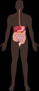
Digestive System: Obtains nutrients from food.
Urinary System: Removes waste and excess water from blood.
Homeostasis
Definition and Importance
Homeostasis is the maintenance of a stable internal environment. Internal conditions are kept within a narrow range, not a fixed state.
Examples of Homeostatic Variables:
Blood pH: 7.35 to 7.45
Internal Body Temperature: 36°C to 37.5°C
Blood Glucose: 70 mg/dL to 90 mg/dL (fasting)
Failure to maintain homeostasis: Results in pathology (disease).
Feedback Loops
Types of Feedback Loops
Feedback loops are mechanisms that control homeostasis:
Negative Feedback: Moves the system back toward the set point; most common; self-regulating.
Positive Feedback: Moves the system away from the set point; less common; requires an 'off switch'.


Negative Feedback Example
Components: Receptor (detects change), Control Center (processes information), Effector (carries out response).
Example: Standing up quickly causes blood pressure to fall; baroreceptors detect this, the medulla signals the heart to increase rate, restoring blood pressure.
Thermoregulation
Receptors: Thermoreceptors in the hypothalamus.
Control Center: Thermoregulatory center in the hypothalamus.
Effectors: Sweat glands (cooling), skeletal muscles (shivering), smooth muscles (control blood flow).


Positive Feedback Example
Blood Clotting: Platelets adhere to wound, release chemicals, attract more platelets, forming a clot.
Labor and Delivery: Baby's head pushes against cervix, pressure signals release of oxytocin, causing contractions, increasing pressure until birth.
Anatomical Position and Directional Terms
Anatomical Position
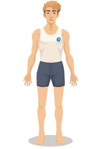
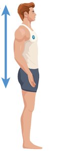
The anatomical position is a universally accepted reference for describing locations and directions on the body:
Body upright, face forward, feet shoulder-width apart, arms at sides, palms forward.
Left and right refer to the subject's left and right.
Directional Terms
Superior: Toward the head
Inferior: Toward the feet
Anterior (Ventral): Toward the front
Posterior (Dorsal): Toward the back
Medial: Toward the midline
Lateral: Away from the midline
Proximal: Closer to the point of attachment (limbs)
Distal: Farther from the point of attachment (limbs)
Superficial: Closer to the surface
Deep: Further from the surface
Anatomical Terms for Body Regions
Head and Neck
Cephalic: Head
Cervical: Neck
Frontal: Forehead
Occipital: Back of the head
Orbital: Eye
Otic: Ear
Buccal: Cheek
Mental: Chin
Trunk
Thoracic: Chest
Abdominal: Abdomen
Umbilical: Navel
Pelvic: Pelvis
Inguinal: Groin
Pubic: Genital region
Back
Scapular: Shoulder blade
Lumbar: Lower back
Sacral: Base of spine
Perineal: Between anus and genitals
Vertebral: Spine
Gluteal: Buttocks
Olecranal: Back of elbow
Arm and Hand
Brachial: Arm
Antebrachial: Forearm
Carpal: Wrist
Pollex: Thumb
Digital: Fingers
Manus: Hand
Antecubital: Front of elbow
Leg and Foot
Femoral: Thigh
Patellar: Knee
Popliteal: Back of knee
Sural: Calf
Crural: Lower leg
Plantar: Sole of foot
Pedal: Foot
Hallux: Big toe
Abdominopelvic Quadrants and Regions
Abdominopelvic Quadrants
Divided into four quadrants: Right Upper (RUQ), Left Upper (LUQ), Right Lower (RLQ), Left Lower (LLQ).
Used to localize pain or pathology.
Example: RLQ pain may indicate appendicitis.
Abdominopelvic Regions
Divided into nine regions: right/left hypochondriac, epigastric, right/left lumbar, umbilical, right/left inguinal, hypogastric.
Used for more precise localization.
Anatomical Planes and Sections
Anatomical Planes
Frontal (Coronal): Divides anterior and posterior.
Sagittal: Divides left and right. Midsagittal is on the midline; parasagittal is off the midline.
Transverse: Divides superior and inferior.
Oblique: Divides at an angle.
Anatomical Sections
Sections are created by cuts along planes for imaging or dissection.
Example: MRI scans can show sagittal, frontal, or transverse sections.
Organization of the Body: Body Cavities
Body Cavities
Anterior (Ventral) Cavity: Houses most organs; divided by the diaphragm into thoracic and abdominopelvic cavities.
Posterior (Dorsal) Cavity: Houses the brain and spinal cord.
Anterior Body Cavity Subdivisions
Thoracic Cavity: Contains heart and lungs; protected by rib cage.
Abdominopelvic Cavity: Contains digestive, urinary, and reproductive organs.
Abdominal Cavity: Most digestive organs and kidneys.
Pelvic Cavity: Bladder and internal reproductive organs.
Thoracic Cavity Organization
Pleural Cavities: Surround the lungs.
Mediastinum: Space between pleural cavities; contains pericardial cavity, esophagus, trachea.
Pericardial Cavity: Surrounds the heart.
Abdominopelvic Cavity Organization
Abdominal Cavity: Liver, digestive organs, pancreas, spleen, kidneys.
Pelvic Cavity: Bladder, internal reproductive organs.
Peritoneal Cavity: Serous membrane-bound; surrounds most abdominal organs.
Retroperitoneum: Space behind peritoneum; houses kidneys, pancreas, rectum.
Organ | Abdominal Cavity | Pelvic Cavity | Peritoneal Cavity |
|---|---|---|---|
Bladder | ✓ | ✓ | |
Stomach | ✓ | ✓ | |
Lungs | |||
Large Intestine | ✓ | ✓ | ✓ |
Kidneys | ✓ |
Additional info: These notes expand on brief points and provide context for exam preparation, including definitions, examples, and logical groupings of anatomical concepts.
