 Back
BackIntroduction to Anatomy and Physiology: The Language, Organization, and Homeostasis of the Human Body
Study Guide - Smart Notes
 Tailored notes based on your materials, expanded with key definitions, examples, and context.
Introduction to Anatomy and Physiology
Overview of Anatomy and Physiology
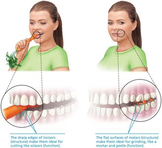
Anatomy and physiology are foundational sciences in understanding the human body. Anatomy is the study of the structure of body parts and their relationships to one another, while physiology focuses on the function of these parts. The principle of 'structure determines function' is central to both fields.
Anatomy: Derived from the Greek meaning 'cutting open'; the oldest form of medical science.
Physiology: Explains how anatomical structures perform their functions.
Example: The sharp edges of incisors (structure) make them ideal for cutting food, while the flat surfaces of molars (structure) are suited for grinding food (function).
Characteristics of Living Things
Properties Shared by All Living Organisms
All living things, including humans, share several essential characteristics that distinguish them from non-living matter.
Cellular Composition: All organisms are composed of cells, the smallest units capable of carrying out life functions.
Movement: Involves contraction of muscle tissue. Skeletal muscle moves body parts, cardiac muscle moves blood, and smooth muscle moves substances like urine and food.
Responsiveness: The ability to detect and respond to stimuli, primarily regulated by the nervous system (e.g., withdrawal reflex from a hot surface).
Excretion: Removal of metabolic wastes, regulated by the digestive, urinary, and respiratory systems.
Reproduction: At the cellular level (cell division for growth and repair) and organismal level (production of offspring, regulated by the endocrine and reproductive systems).
Growth: Increase in size of a body part or the organism as a whole.
Metabolism: The sum of all chemical reactions in the body, including catabolism (breaking down substances) and anabolism (synthesizing substances). Cellular respiration produces ATP for cellular work.

Levels of Organization in the Human Body
Hierarchical Structure of the Body
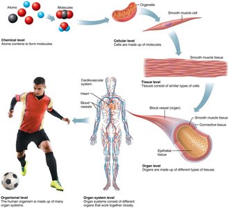
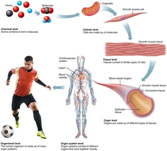
The human body is organized into a hierarchy of structural levels, each building upon the previous one. This organization is essential for understanding how the body functions as a whole.
Chemical Level: Atoms combine to form molecules.
Cellular Level: Molecules form organelles, which make up cells.
Tissue Level: Groups of similar cells form tissues.
Organ Level: Two or more tissues form organs.
Organ System Level: Groups of organs work together to perform complex functions.
Organismal Level: All organ systems working together constitute the living organism.
Major Organ Systems
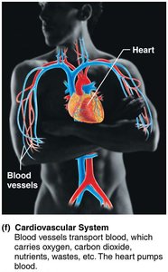
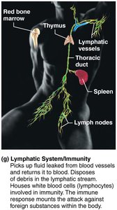
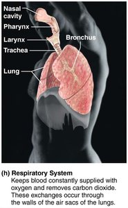
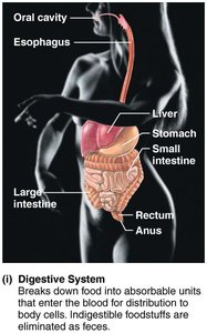
The human body contains 11 major organ systems, each with specific organs and functions. These systems work together to maintain life and homeostasis.
System | Major Organs | Functions |
|---|---|---|
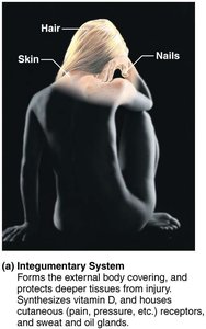
Integumentary | Skin, hair, nails, sweat glands | Protection, temperature regulation, sensory information |
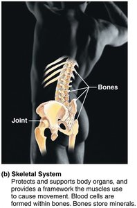
Skeletal | Bones, cartilages, ligaments | Support, protection, mineral storage, blood cell formation |
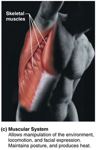
Muscular | Skeletal muscles, tendons | Movement, posture, heat production |
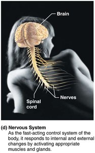
Nervous | Brain, spinal cord, nerves, sense organs | Control, coordination, response to stimuli |
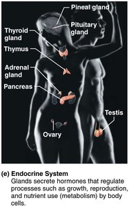
Endocrine | Glands (thyroid, pancreas, etc.) | Hormone production, regulation of metabolism and growth |
Cardiovascular | Heart, blood, blood vessels | Transport of substances, heat distribution |
Lymphatic | Spleen, thymus, lymph nodes, vessels | Immunity, fluid return to blood |
Respiratory | Lungs, trachea, bronchi, alveoli | Gas exchange |
Digestive | Stomach, intestines, esophagus, teeth | Breakdown and absorption of nutrients |
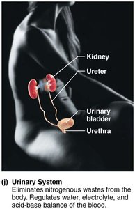
Urinary | Kidneys, ureters, bladder, urethra | Excretion, water and pH balance |
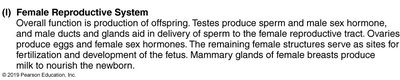
Reproductive | Male: testes, penis, prostate; Female: ovaries, uterus, vagina | Production of offspring |

Anatomical Position and Directional Terms
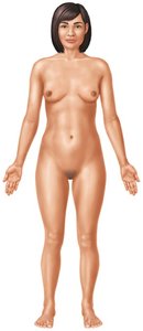
Anatomical Position
The anatomical position is a standardized posture used as a reference in describing the location and relation of body parts. The body stands upright, facing forward, with feet shoulder-width apart, arms at the sides, and palms facing forward.
Directional Terms
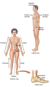
Directional terms describe the positions of structures relative to other structures or locations in the body. They are often used in pairs with opposite meanings.
Superior (cranial): Toward the head or upper part of a structure.
Inferior (caudal): Away from the head or toward the lower part of a structure.
Anterior (ventral): Toward the front of the body.
Posterior (dorsal): Toward the back of the body.
Medial: Toward the midline of the body.
Lateral: Away from the midline of the body.
Proximal: Closer to the point of attachment or origin.
Distal: Farther from the point of attachment or origin.
Superficial (external): Toward or on the surface of the body.
Deep (internal): Away from the surface of the body.
Planes and Sections of the Body
Body Planes
Body planes are imaginary flat surfaces that divide the body into sections. They are used to describe locations and directions in anatomy.
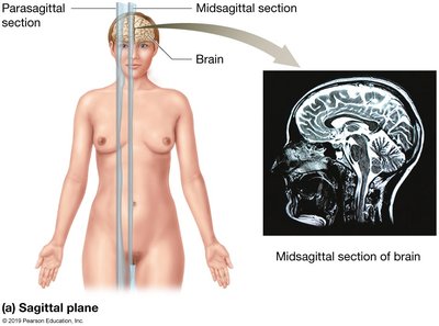
Sagittal Plane: Divides the body into left and right portions. A midsagittal plane divides the body into equal left and right halves, while a parasagittal plane divides it into unequal portions.
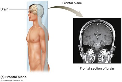
Frontal (Coronal) Plane: Divides the body into anterior (front) and posterior (back) portions.
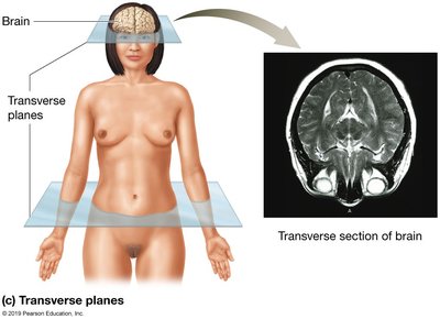
Transverse (Horizontal) Plane: Divides the body into superior (upper) and inferior (lower) portions.
Body Cavities
Major Body Cavities
Body cavities are spaces within the body that protect, separate, and support internal organs. The body is divided into axial (head, neck, trunk) and appendicular (limbs) portions. The axial portion contains the main body cavities:
Posterior (Dorsal) Cavity: Protects the nervous system; includes the cranial cavity (brain) and vertebral cavity (spinal cord).
Anterior (Ventral) Cavity: Houses internal organs; includes the thoracic cavity (heart, lungs) and abdominopelvic cavity (digestive, urinary, reproductive organs).

Thoracic and Abdominopelvic Cavities
Thoracic Cavity: Contains pleural cavities (lungs), mediastinum (heart, trachea, esophagus), and pericardial cavity (heart).
Abdominopelvic Cavity: Extends from the diaphragm to the pelvis; includes abdominal cavity (stomach, liver, intestines) and pelvic cavity (bladder, reproductive organs).

Abdominopelvic Quadrants and Regions
The abdominopelvic cavity is further divided for clinical and anatomical reference:
Four Quadrants: Right upper, left upper, right lower, left lower (centered on the umbilicus).
Nine Regions: A tic-tac-toe grid for more precise localization (e.g., epigastric, hypogastric, lumbar regions).
Serous Membranes
Membranes of Body Cavities
Serous membranes line the walls and organs of the thoracic and abdominopelvic cavities, reducing friction and compartmentalizing organs.
Pleural Membranes: Parietal pleura lines the thoracic cavity; visceral pleura covers the lungs.
Pericardial Membranes: Parietal pericardium lines the pericardial cavity; visceral pericardium covers the heart.
Peritoneal Membranes: Parietal peritoneum lines the abdominopelvic cavity; visceral peritoneum covers abdominal organs. Some organs (e.g., kidneys) are retroperitoneal (behind the peritoneum).
Regional Terms
Body Regions and Landmarks
Regional terms are used to specify locations on the body, such as cephalic (head), cervical (neck), thoracic (chest), brachial (arm), and femoral (thigh). These terms are essential for precise anatomical communication.
Homeostasis
Definition and Importance
Homeostasis is the maintenance of a stable internal environment, essential for survival. The body's internal environment includes interstitial fluid, blood plasma, cerebrospinal fluid, and more. Physiological systems constantly monitor and adjust to maintain homeostasis; failure leads to disease or death.
Control of Homeostasis: Feedback Loops
Homeostasis is maintained through feedback loops involving three main components:
Receptor: Detects changes (stimuli) and sends information to the control center.
Control Center: Sets the range for the variable, evaluates input, and generates output commands.
Effector: Receives output and produces a response to change the variable, opposing or enhancing the original stimulus.
There are two main types of feedback loops:
Negative Feedback: The response reduces or shuts off the original stimulus, returning the variable to its normal range (e.g., body temperature regulation).
Positive Feedback: The response enhances or exaggerates the original stimulus, moving the variable further from its original value (e.g., blood clotting).
Example of Negative Feedback: Regulation of body temperature. If body temperature rises, mechanisms such as sweating are activated to cool the body.
Example of Positive Feedback: Blood clotting, where each step accelerates the process until the clot is formed.
Additional info: Homeostatic mechanisms are crucial for maintaining variables such as blood pressure, heart rate, and electrolyte concentrations within narrow limits.
