 Back
Introduction to Human Anatomy and Physiology: Concepts, Terminology, and Tissue Organization
Study Guide - Smart Notes
 Tailored notes based on your materials, expanded with key definitions, examples, and context.
Concepts & Terminology in Anatomy and Physiology
Definitions and Scope
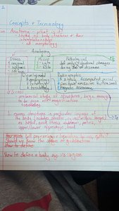
Anatomy and physiology are foundational sciences in understanding the structure and function of the human body. Anatomy focuses on the structure of body parts, while physiology explores their functions and how they work together to sustain life.
Anatomy: The study of the structure of body parts and their relationships to one another.
Physiology: The study of the function of the body’s structural machinery.
Subdivisions of Anatomy:
Gross (Macroscopic) Anatomy: Study of large body structures visible to the naked eye (e.g., heart, lungs, kidneys).
Microscopic Anatomy: Study of structures too small to be seen with the naked eye (e.g., cells, tissues).
Developmental Anatomy: Study of structural changes throughout the lifespan.
Subdivisions of Physiology:
Renal Physiology: Concerns kidney function and urine production.
Neurophysiology: Explains the workings of the nervous system.
Cardiovascular Physiology: Examines the operation of the heart and blood vessels.
Levels of Structural Organization
Hierarchy of Organization
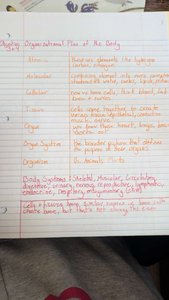
The human body is organized into a hierarchy of structural levels, each building on the previous one. Understanding these levels is essential for grasping how the body functions as a whole.
Chemical Level: Atoms combine to form molecules.
Cellular Level: Cells are made up of molecules.
Tissue Level: Tissues consist of similar types of cells.
Organ Level: Organs are made up of different types of tissues.
Organ System Level: Organ systems consist of different organs that work closely together.
Organismal Level: The human organism is made up of many organ systems.
Homeostasis and Control Mechanisms
Definition and Importance
Homeostasis is the maintenance of a stable internal environment despite changes in the external environment. It is vital for normal body functioning and sustaining life.
Components of Homeostatic Control:
Receptor: Detects changes (stimuli) and sends information to the control center.
Control Center: Determines the set point and analyzes input; sends output to the effector.
Effector: Carries out the control center’s response to the stimulus.
Feedback Mechanisms:
Negative Feedback: Reduces or shuts off the original stimulus (e.g., regulation of body temperature, blood glucose levels).
Positive Feedback: Enhances the original stimulus (e.g., blood clotting, labor contractions).

Body Cavities and Serous Membranes
Major Body Cavities
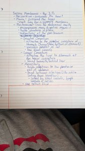
The body contains several cavities that house and protect vital organs. These cavities are lined by serous membranes that reduce friction between organs.
Dorsal Body Cavity: Protects the nervous system and is divided into the cranial and vertebral cavities.
Ventral Body Cavity: Houses the thoracic and abdominopelvic cavities.
Serous Membranes
Parietal Serosa: Lines internal body cavity walls.
Visceral Serosa: Covers internal organs.
Serous Fluid: Separates the serosae, reducing friction.
Abdominopelvic Regions and Quadrants
Division of the Abdomen
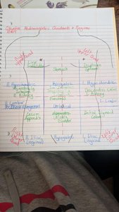
The abdominopelvic cavity is divided into regions and quadrants for anatomical reference and clinical assessment.
Four Quadrants: Right Upper, Left Upper, Right Lower, Left Lower.
Nine Regions: Right/Left Hypochondriac, Epigastric, Right/Left Lumbar, Umbilical, Right/Left Iliac (Inguinal), Hypogastric (Pubic).
Microscopy and Histology
Microscopic Anatomy
Microscopy is essential for studying structures that cannot be seen with the naked eye, such as cells and tissues. Histology is the study of tissues, which are groups of similar cells performing a common function.
Types of Tissues: Epithelial, Connective, Muscle, Nervous.
Histological Techniques: Involve preparing tissue sections for microscopic examination.
Epithelial Tissue
Classification and Function
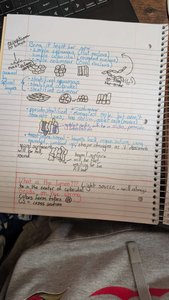
Epithelial tissues cover body surfaces, line cavities, and form glands. They are classified based on cell shape and number of layers.
Shapes: Squamous (flat), Cuboidal (cube-shaped), Columnar (tall).
Layers: Simple (one layer), Stratified (multiple layers).
Functions: Protection, absorption, filtration, secretion.
Connective Tissue
Structure and Types
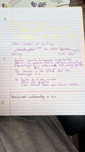
Connective tissue supports, protects, and binds other tissues together. It consists of cells, fibers, and ground substance (extracellular matrix).
Main Types:
Connective Tissue Proper (Loose and Dense)
Cartilage
Bone
Blood
Functions: Support, protection, insulation, transportation of substances.
Summary Table: Levels of Organization
Level | Description |
|---|---|
Chemical | Atoms and molecules |
Cellular | Cells and their organelles |
Tissue | Groups of similar cells |
Organ | Contains two or more types of tissues |
Organ System | Organs that work closely together |
Organismal | All organ systems combined |
Key Concepts and Applications
Homeostasis: Maintained by negative and positive feedback mechanisms.
Body Cavities: Provide protection and allow organ movement.
Tissues: Four basic types, each with specialized functions.
Example: Blood glucose regulation is a classic example of negative feedback, where insulin and glucagon maintain glucose homeostasis.
Additional info: These notes provide foundational knowledge for further study in anatomy and physiology, including the structure and function of cells, tissues, and organ systems.
