 Back
BackIntroduction to Human Anatomy: Study Guide for Lab Exam 1
Study Guide - Smart Notes
 Tailored notes based on your materials, expanded with key definitions, examples, and context.
Introduction to Anatomy
Directional Terms
Directional terms are used to describe the locations of structures in relation to other structures or locations in the body. Mastery of these terms is essential for clear anatomical communication.
Superior (Cranial): Toward the head or upper part of a structure (e.g., the heart is superior to the liver).
Inferior (Caudal): Away from the head or toward the lower part of a structure (e.g., the stomach is inferior to the lungs).
Anterior (Ventral): Toward or at the front of the body (e.g., the sternum is anterior to the heart).
Posterior (Dorsal): Toward or at the back of the body (e.g., the spine is posterior to the stomach).
Medial: Toward the midline of the body (e.g., the nose is medial to the eyes).
Lateral: Away from the midline of the body (e.g., the arms are lateral to the chest).
Proximal: Closer to the origin of a body part or the point of attachment (e.g., the elbow is proximal to the wrist).
Distal: Farther from the origin of a body part (e.g., the fingers are distal to the elbow).
Superficial: Toward or at the body surface (e.g., the skin is superficial to the muscles).
Deep: Away from the body surface; more internal (e.g., the lungs are deep to the rib cage).
Body Cavities
The body contains several cavities that house and protect vital organs:
Cranial cavity: Contains the brain.
Vertebral (spinal) cavity: Contains the spinal cord.
Thoracic cavity: Contains the heart and lungs.
Abdominopelvic cavity: Subdivided into the abdominal cavity (digestive organs, spleen, kidneys) and pelvic cavity (bladder, reproductive organs, rectum).
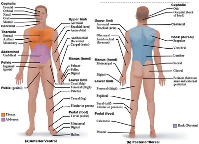
Anatomical Areas of the Body
Specific regions of the body are identified by anatomical terms. These terms are used to describe locations for clinical and educational purposes.
Sternal: Area of the sternum (breastbone).
Axillary: Armpit region.
Brachial: Arm region.
Femoral: Thigh region.
Patellar: Anterior knee (kneecap) region.
Other regions: See image below for a comprehensive overview.
Planes of the Body
Body planes are imaginary lines used to divide the body into sections for anatomical study:
Frontal (coronal) plane: Divides the body into anterior and posterior parts.
Sagittal plane: Divides the body into right and left parts (midsagittal is exactly in the midline; parasagittal is off-center).
Transverse (horizontal) plane: Divides the body into superior and inferior parts.
Serous Membranes
Serous membranes (serosa) are thin, double-layered membranes that cover organs and line body cavities. They reduce friction between moving organs.
Pericardium: Surrounds the heart.
Pleura: Surrounds the lungs.
Peritoneum: Surrounds abdominal organs.
Serosa: General term for serous membranes.
Abdominopelvic Quadrants and Regions
The abdominopelvic area is divided for clinical reference:
Four quadrants: Right upper, left upper, right lower, left lower.
Nine regions: Right hypochondriac, epigastric, left hypochondriac, right lumbar, umbilical, left lumbar, right iliac (inguinal), hypogastric (pubic), left iliac (inguinal).
Organs in each region: For example, the liver is in the right hypochondriac and epigastric regions; the appendix is in the right iliac region.
Anatomical Position
The anatomical position is the standard reference for describing locations and directions in the body:
Body is erect, facing forward.
Arms at sides, palms facing forward.
Feet slightly apart.
Organ System Overview
The 11 Major Organ Systems
The human body is organized into 11 major organ systems, each with specific functions and major organs:
Integumentary System: Skin, hair, nails; protects body, synthesizes vitamin D, houses cutaneous receptors.
Skeletal System: Bones, joints; supports and protects organs, provides framework for muscles, forms blood cells.
Muscular System: Skeletal muscles; allows movement, maintains posture, produces heat.
Nervous System: Brain, spinal cord, nerves; fast-acting control system, responds to stimuli.
Endocrine System: Glands (pituitary, thyroid, etc.); secretes hormones for regulation of growth, metabolism, reproduction.
Cardiovascular System: Heart, blood vessels; transports blood, nutrients, gases, wastes.
Lymphatic System: Lymph nodes, lymphatic vessels, spleen; returns fluid to blood, defends against pathogens.
Respiratory System: Lungs, trachea, bronchi; supplies blood with oxygen, removes carbon dioxide.
Digestive System: Stomach, intestines, liver, pancreas; breaks down food, absorbs nutrients, eliminates waste.
Urinary System: Kidneys, bladder, urethra; eliminates nitrogenous wastes, regulates water and electrolytes.
Reproductive System: Testes, ovaries, uterus; produces offspring.
Organ Identification in Lab and Images
Be able to identify major organs and anatomical regions on diagrams, models, and images, including those from fetal pig dissections and PowerPoint slides. Practice labeling and recognizing these structures for exam preparation.
Mastering A&P – Organ Systems
Review and practice labeling all images and diagrams provided in the Mastering A&P assignments, focusing on the identification of organs and their associated systems.
Additional info: This guide expands on the provided study list with academic definitions, examples, and context for each topic. The included image directly supports the section on anatomical regions and terminology.
