 Back
BackChapter 1
Study Guide - Smart Notes
 Tailored notes based on your materials, expanded with key definitions, examples, and context.
Scope of Microbiology
Introduction
Microbiology is the study of microscopic organisms, including bacteria, viruses, fungi, and protozoa. This field explores the structure, function, classification, and significance of microorganisms in health, disease, and the environment.
Achievements of Early Microbiologists
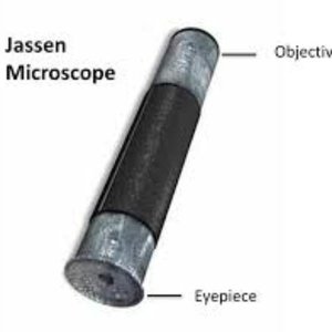
Zaccharias and Hans Janssen
Invention of the Compound Microscope: Dutch eyeglass makers who created the first compound microscope, consisting of a simple tube with lenses at each end.
Magnification: Their device could magnify objects between 3x and 9x, depending on the light source.
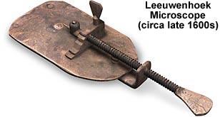
Antony van Leeuwenhoek
Father of Microscopy: First to observe live bacteria and protozoans using a simple, single-lens microscope.
Discovery of Animalcules: Described small living organisms as "animalcules," laying the foundation for microbiology.
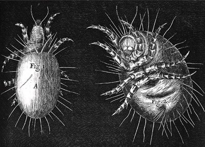
Robert Hooke
Improvements to Microscopy: Enhanced the compound light microscope and observed various biological specimens, including plant cells and insects.
Publication: Authored Micrographia, which included detailed illustrations of microscopic observations.
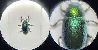
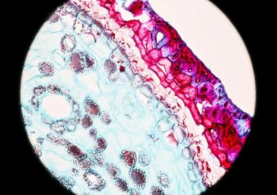
Types of Microscopes and Their Uses
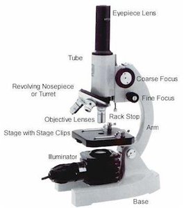
Light Microscopes
Light microscopes use visible light and optical lenses to magnify specimens. They are classified as simple (one lens) or compound (multiple lenses).
Ocular Lens: Located in the eyepiece.
Objective Lens: Located in the body of the microscope.
Total Magnification: Calculated as the product of the ocular and objective lens magnifications (e.g., 10x ocular × 4x objective = 40x).
Stereomicroscope (Dissecting Microscope)
Low Magnification: Used for viewing larger, three-dimensional objects, often during dissection.
Three-Dimensional Imaging: Provides depth perception for examining specimen surfaces.
Specialized Light Microscopy Techniques
Bright-Field: Produces a bright background; specimens often require staining.
Dark-Field: Illuminates specimen edges against a dark background; useful for observing motility and hard-to-stain organisms.
Phase-Contrast: Enhances contrast in transparent specimens by exploiting differences in refractive index.
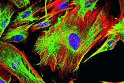
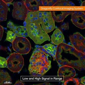
Fluorescence: Uses UV light to excite fluorescent dyes in specimens, commonly used in diagnostics.
Confocal: Uses lasers to produce sharp, high-resolution images of thick specimens.
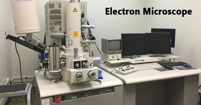
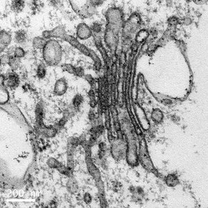
Electron Microscopes
Electron microscopes use electron beams and magnetic fields for imaging, offering much higher resolution than light microscopes.
Transmission Electron Microscope (TEM): Electrons pass through ultra-thin specimens, producing detailed two-dimensional images. Magnification up to 1,000,000x.
Scanning Electron Microscope (SEM): Scans the surface of specimens, creating three-dimensional images with a large depth of field. Magnification up to 100,000x.
Theory of Spontaneous Generation
Historical Experiments
Francesco Redi (1668): Demonstrated that maggots arise from fly eggs, not spontaneously from meat.
John Needham (1745): Claimed spontaneous generation after observing microbial growth in boiled broth.
Lazzaro Spallanzani: Showed that boiling broth in sealed flasks prevented microbial growth, suggesting microbes come from the air.
Louis Pasteur (1861): Used swan-neck flasks to prove that microorganisms in the air, not spontaneous generation, cause contamination.
John Tyndall & Ferdinand Cohn: Provided evidence for heat-resistant bacteria and bacterial endospores.
Pasteurization
Developed by Louis Pasteur: Heating liquids to 55°C for several minutes reduces microbial load, preventing spoilage and disease.
Sterilization: Complete destruction of all microorganisms and endospores.
Germ Theory of Disease
Significance and Key Contributors
Oliver Wendell Holmes & Ignaz Semmelweis: Demonstrated the importance of hand hygiene in preventing disease transmission.
Joseph Lister: Introduced aseptic techniques and carbolic acid to reduce surgical infections.
Louis Pasteur & Robert Koch: Established that specific microbes cause specific diseases.
Edward Jenner: Developed the first vaccine (smallpox) using cowpox virus.
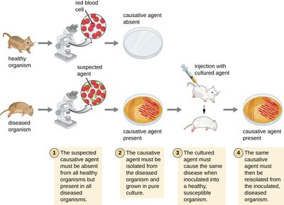
Koch’s Postulates
Steps to Identify Disease-Causing Microbes
The suspected pathogen must be present in all cases of the disease and absent from healthy organisms.
The pathogen must be isolated and grown in pure culture.
The cultured pathogen must cause the same disease when introduced into a healthy host.
The pathogen must be re-isolated from the experimentally infected host and identified as the same organism.
Origin and Evolution of Microorganisms
Timeline and Evolutionary Relationships
Earth’s Formation: Approximately 4.5 billion years ago.
First Life Forms: Prokaryotes appeared between 3.5 and 4 billion years ago.
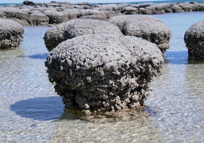
Stromatolites: Fossilized microbial mats, evidence of ancient life.
Evolution: Gradual changes over millions of years led to the diversity of life, including the emergence of eukaryotes from prokaryotes.
Domains of Life: Bacteria, Archaea, and Eukarya, determined by nucleic acid sequencing.
Differences Between Prokaryotes and Eukaryotes
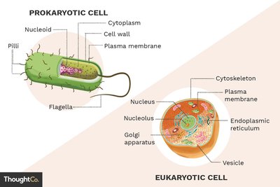
Cellular Organization
Prokaryotes: Lack a nucleus and membrane-bound organelles; include Bacteria and Archaea.
Eukaryotes: Possess a nucleus and membrane-bound organelles; include algae, fungi, and protozoans.
Viruses: Non-cellular, require host cells for replication.
Prions and Viroids: Infectious agents lacking cellular structure; prions are misfolded proteins, viroids are plant pathogens composed of RNA.
Role of Taxonomy
Classification and Nomenclature
Taxonomy: The science of organizing, classifying, and naming living organisms.
Taxa: Non-overlapping groups based on similarities.
Nomenclature: Binomial system (Genus species), e.g., Homo sapiens.
Identification: Determining and recording traits of organisms.
Microbial Transmission
Modes of Transmission
Biofilms: Communities of microbes living within a polymeric matrix on surfaces.
Relationships: Mutualism, commensalism, synergism, and parasitism.
Transmission Routes:
Waterborne: Through contaminated water.
Foodborne: Through contaminated food.
Airborne: Via aerosols from coughing, sneezing, or talking.
Zoonotic: From animals to humans.
Uses of Microorganisms in Everyday Life
Applications
Food Production: Fermentation processes for vinegar, yogurt, cheese, bread, and alcoholic beverages.
Water Treatment: Microbial analysis and purification.
Pharmaceuticals: Production of antibiotics, hormones, and other drugs.
Agriculture: Soil fertility, plant disease management, and crop growth.
Bioremediation: Use of microbes to degrade environmental pollutants.
Energy: Bioconversion of organic material into fuels like methane.
Forensics: Microbial analysis in crime and outbreak investigations.
