 Back
BackIntroduction to Prokaryotes: Structure, Function, and Clinical Relevance
Study Guide - Smart Notes
 Tailored notes based on your materials, expanded with key definitions, examples, and context.
Introduction to Prokaryotes
Overview of Prokaryotic Domains
Prokaryotes are unicellular organisms that lack a membrane-bound nucleus and organelles. They are classified into two domains: Bacteria and Archaea. Understanding the differences and similarities between these domains is fundamental for microbiology and clinical practice.
Bacteria and Archaea share a common ancestor but differ in cell wall composition, membrane lipids, and genetic machinery.
Both are distinct from Eukarya, which includes all eukaryotic organisms.

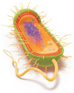
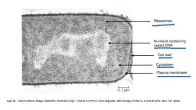
Basic Structure of Prokaryotic Cells
Prokaryotic cells are structurally simpler than eukaryotic cells but possess specialized features for survival and adaptation.
Unicellular and lack a membrane-bound nucleus.
Genetic material is located in the nucleoid region.
Most have a cell wall for structural support and protection.
Other features include ribosomes, plasma membrane, capsule (in some), fimbriae, and flagella (in some).
Prokaryotic Cell Morphology
Sizes and Surface Area-to-Volume Ratio
Prokaryotic cells are generally small, ranging from 0.2 to 750 μm in diameter, with most between 0.5 and 2.0 μm. Their small size is due to reliance on diffusion for nutrient uptake, which is limited by the surface area-to-volume ratio.
Smaller cells have a higher surface area-to-volume ratio, facilitating efficient nutrient and waste exchange.
Examples: Mycoplasma species are among the smallest; Thiomargarita namibiensis is among the largest.

Shapes and Arrangements
Prokaryotes exhibit diverse shapes and arrangements, which are important for identification and pathogenicity.
Bacilli: Rod-shaped
Cocci: Spherical
Vibrio: Comma-shaped
Stella: Star-shaped
Coccobacilli: Ovoid
Spirochetes: Spiral-shaped
Pleomorphic: Variable shapes, enhancing survival and transmission
Cell Arrangements
Arrangements result from cell division patterns:
Diplococci: Pairs of cocci
Streptococci: Chains of cocci
Staphylococci: Clusters of cocci
Diplobacilli: Pairs of bacilli
Streptobacilli: Chains of bacilli
Palisades: Clusters of bacilli

Pleomorphism
Pleomorphic bacteria can change shape, which may enhance their ability to evade the immune system and adapt to different environments, impacting their pathogenicity.
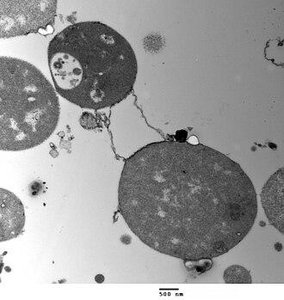
Prokaryotic Cell Division
Binary Fission
Prokaryotic cells primarily reproduce by binary fission, an asexual process:
DNA is replicated.
Cell elongates.
Chromosomes are segregated to opposite ends.
A septum forms at the midpoint.
Two genetically identical daughter cells are produced.
Prokaryotic Cell Envelope
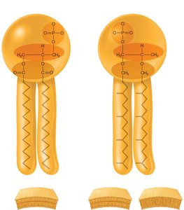
Plasma Membrane Structure and Function
The plasma membrane is a thin, flexible phospholipid bilayer that acts as a selective barrier, controlling the movement of substances in and out of the cell.
Composed of lipids and proteins.
Proteins serve as transporters, anchors, receptors, and enzymes.
Site of metabolic reactions, including ATP synthesis.

Membrane Fluidity
Membrane fluidity is essential for cell function and is influenced by temperature and fatty acid composition:
Warmer temperatures increase fluidity; colder temperatures decrease it.
Unsaturated fatty acids enhance fluidity; saturated fatty acids make the membrane more rigid.
Differences Between Bacterial and Archaeal Membranes
Bacteria: Linear fatty acids, ester bonds, always bilayers.
Archaea: Branched isoprene chains, ether bonds, can form monolayers or bilayers (especially in extreme environments).
Cell Wall Structure and Function
The cell wall provides rigidity and protection. Its composition differs between Bacteria and Archaea:
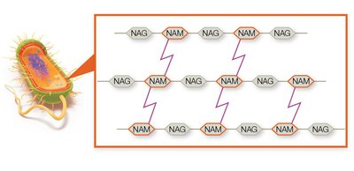
Bacteria: Peptidoglycan (alternating N-acetylglucosamine [NAG] and N-acetylmuramic acid [NAM] with peptide cross-links).
Archaea: Pseudopeptidoglycan or other polymers.
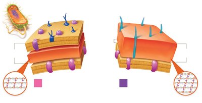
Gram-Positive vs. Gram-Negative Cell Walls
Gram staining differentiates bacteria based on cell wall structure:
Gram-positive: Thick peptidoglycan layer, teichoic acids, no outer membrane, stains purple.
Gram-negative: Thin peptidoglycan layer, outer membrane with lipopolysaccharide (LPS), porins, stains pink/red.
Cell Feature | Gram-Negative | Gram-Positive |
|---|---|---|
Outer membrane | Yes | No |
Lipid A (endotoxin) | Yes | No |
Porins | Yes | No |
Teichoic acids | No | Yes |
Peptidoglycan | Thin | Thick |
Gram stain color | Pink | Purple |
Resistance to drying | No | Yes |
Penicillin susceptibility | Low | High |
Clinical Implications of Gram Status
Gram-negative bacteria are harder to kill due to the outer membrane and can release endotoxins (Lipid A) that trigger strong immune responses.
Gram-positive bacteria are more susceptible to antibiotics targeting peptidoglycan but can survive longer on surfaces.
Acid-Fast Bacteria
Some bacteria (e.g., Mycobacterium, Nocardia) have waxy cell walls containing mycolic acid, making them resistant to Gram staining. Acid-fast staining is used for their identification.
Acid-fast bacteria appear red/pink after staining.
Clinically important for diagnosing tuberculosis and leprosy.
Mycoplasma and L-Forms
Mycoplasma: Lack a cell wall, have sterol-rich membranes, are pleomorphic, and often live inside host cells.
L-forms: Bacteria that have lost their cell wall, can persist in the host, and are resistant to antibiotics targeting cell walls.
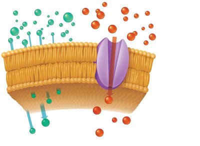
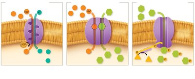
Transport Mechanisms in Prokaryotes
Passive Transport
Simple diffusion: Movement of small, noncharged molecules down their concentration gradient.
Facilitated diffusion: Movement of substances via membrane proteins (channels or carriers) down their concentration gradient.
Osmosis
Osmosis is the diffusion of water across a selectively permeable membrane from low to high solute concentration.
Isotonic: No net water movement.
Hypertonic: Water leaves the cell, causing plasmolysis.
Hypotonic: Water enters the cell, causing swelling or lysis if the cell wall is damaged.
Active Transport
Primary active transport: Uses ATP to move substances against their gradient.
Secondary active transport: Uses ion gradients (symport or antiport).
Phosphotransferase system: Chemically modifies and transports substances (e.g., glucose).
Extracellular Structures
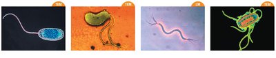
Flagella
Flagella are long, whip-like structures used for motility. They are composed of flagellin and anchored differently in Gram-positive (2 rings) and Gram-negative (4 rings) bacteria.
Enable movement via a run-and-tumble mechanism.
Types of taxis: chemotaxis (chemicals), phototaxis (light), aerotaxis (oxygen).
Arrangements: monotrichous, lophotrichous, amphitrichous, peritrichous.
Fimbriae and Pili
Fimbriae: Short, bristle-like structures for adhesion and biofilm formation.
Pili: Longer, less numerous, involved in adhesion, movement, and gene transfer (conjugation).
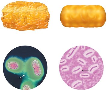
Glycocalyx
Sticky, carbohydrate-rich layer outside the cell wall.
Types: Slime layer (loose, unorganized) and capsule (tight, organized).
Functions: adhesion, protection from desiccation, immune evasion.
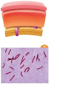
Biofilms
Biofilms are structured communities of bacteria adhering to surfaces and embedded in a self-produced matrix. They are highly resistant to antibiotics and immune responses.
Common on teeth (dental plaque), medical devices, and tissues.
Responsible for 60–80% of human infections.
Intracellular Structures
Nucleoid and Plasmids
Nucleoid: Region containing the single, circular chromosome.
Plasmids: Small, circular DNA molecules carrying non-essential but advantageous genes (e.g., antibiotic resistance).
Ribosomes
Sites of protein synthesis, composed of RNA and protein.
Prokaryotic ribosomes are 70S (50S + 30S subunits).
Cytoskeleton
Composed of protein filaments, provides structure and support.
Inclusion Bodies
Storage sites for nutrients and other substances (e.g., carboxysomes, magnetosomes).
Endospores
Endospores are dormant, highly resistant structures formed by certain bacteria (e.g., Bacillus, Clostridium) to survive harsh conditions.
Resistant to heat, drying, radiation, and disinfectants.
Pose challenges in healthcare and food safety due to their resilience.
Sterilization requires autoclaving.
Sporulation
Process of endospore formation: DNA replication, packaging, formation of protective layers, release.
Endospores germinate into vegetative cells when conditions improve.
Summary Table: Key Differences Between Prokaryotic Cell Types
Feature | Bacteria | Archaea | Eukarya |
|---|---|---|---|
Nucleus | No | No | Yes |
Cell Wall | Peptidoglycan | Pseudopeptidoglycan/other | Cellulose/chitin/none |
Membrane Lipids | Ester-linked, unbranched | Ether-linked, branched | Ester-linked, unbranched |
Ribosomes | 70S | 70S | 80S |
Reproduction | Binary fission | Binary fission | Mitosis/meiosis |
Additional info: This guide integrates foundational microbiology with clinical implications, supporting ANP college-level understanding of prokaryotic cell structure, function, and relevance to health sciences.
